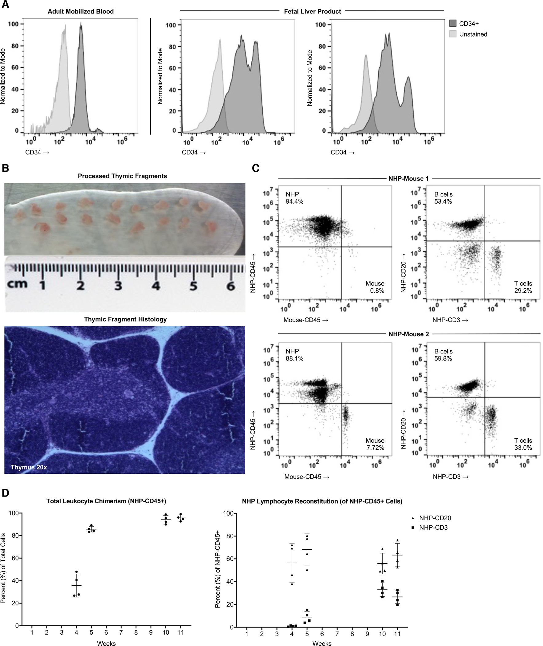
Figure 2. Robust Engraftment of Fetal Liver HSPCs and Thymic Tissue in NOG-EXL Mice.

(A) NHP-CD34 hematopoietic stem and progenitor cell (HSPC) purity was measured following MACS-purification of rhesus mobilized blood (left, negative control = unstained cells) and fetal liver (right, negative control = negative MACS fraction), including CD34lo and CD34hi populations. Mobilized blood was from the same donor but a separate purification than shown in Figure 1. (B) Thymus tissue was dissected into 1mm × 1mm fragments suitable for primatization experiments, and histological analysis of H&E stained thymus sections shows anatomical structures required for T cell development (medulla, cortex, Hassell’s corpuscles). (C) Two NOG-EXL mice were transplanted with a common source of fetal rhesus thymus tissue and IV injected with 1×10⁶ CD34+ cells isolated from fetal liver of the same donor. Overall engraftment (NHP-CD45 vs mouse-CD45) at week 11 post-surgery is shown on the left, and the proportion of the NHP-CD45 cells expressing the B cell marker CD20 and T cell marker CD3 is shown on the right. (D) Engraftment kinetics are shown from week 4 post-primatization surgery to week 11 in a cohort of 6 mice made from a second tissue source, started at the same time. Two mice were analyzed at all timepoints, two were removed after 5 weeks due to premature death, and two were included in the analysis at 10 and 11 weeks (logistical issues preclude their analyses at early timepoints).
