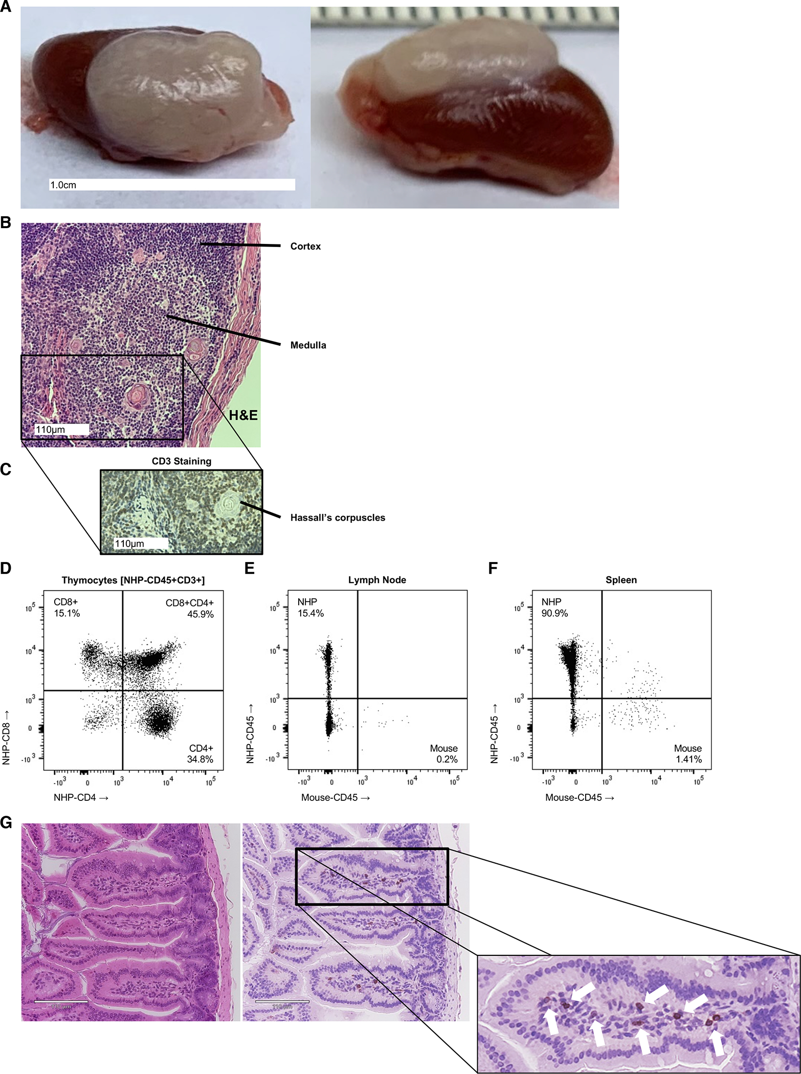
Figure 4. Fetal Thymic Organoid, Secondary Lymphoid and Tissue-Resident Immune Cells.

Thymic organoids formed in engrafted NHP mice (A) shown grossly, with (B) H&E staining including well-organized medullary and cortical regions, as well as Hassall’s corpuscles, consistent with native thymic histology. (C) Anti-CD3 (rhesus) staining identifies the presence of diffuse T cell distribution within the cortex and medulla, which is supported by (D) flow cytometric analysis of CD4+ and CD8+, including developing double positive CD4+CD8+ subsets within the thymic organoid of a fully engrafted primatized mouse. Secondary lymphoid tissue, including lymph node (E) and spleen (F) are populated with NHP-derived leukocytes (NHP-CD45+) as demonstrated by flow cytometric analysis of NHP-CD45+ staining. (G) Cross-sectional H&E and anti-CD3 stained histology reveals tissue-resident T and non-T lymphocytes (denoted by arrows) within the intestinal villi of the mature NHP mouse.
