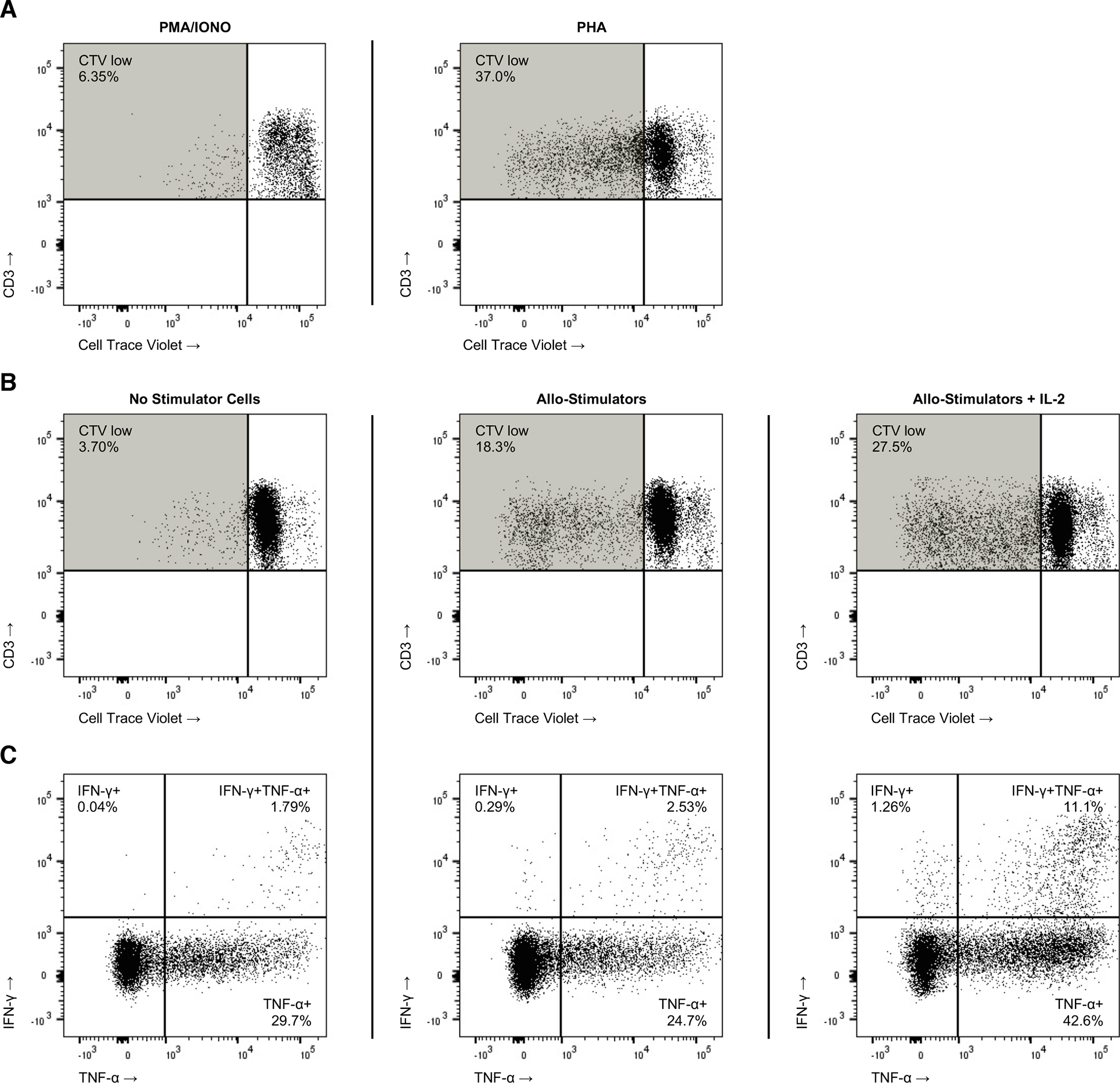
Figure 6. NHP Mice Immune Cell Function.

Splenocytes from primary NHP mice were harvested, labeled with Cell Trace Violet (CTV), and then stimulated with 10x PMA/Ionomycin or PHA (10ug/ml), which (A) resulted in robust T cell proliferation [CTV(lo)] in culture. A mixed lymphocyte reaction (MLR) was performed with isolated NHP mouse splenocytes mixed in a 2:1 dilution with counter-labeled, irradiated adult NHP PBMC (B) yielding multiple cycles of in vitro T cell proliferation in response to allogenic stimulation; IL-2 (100ng) was added for one condition as an additional inflammatory stimulus present in the post-transplantation microenvironment. At the termination of the MLR, cells were additionally assayed (C) by intracellular flow cytometry for interferon gamma (IFNγ) and tumor necrosis factor alpha (TNFα) production.
