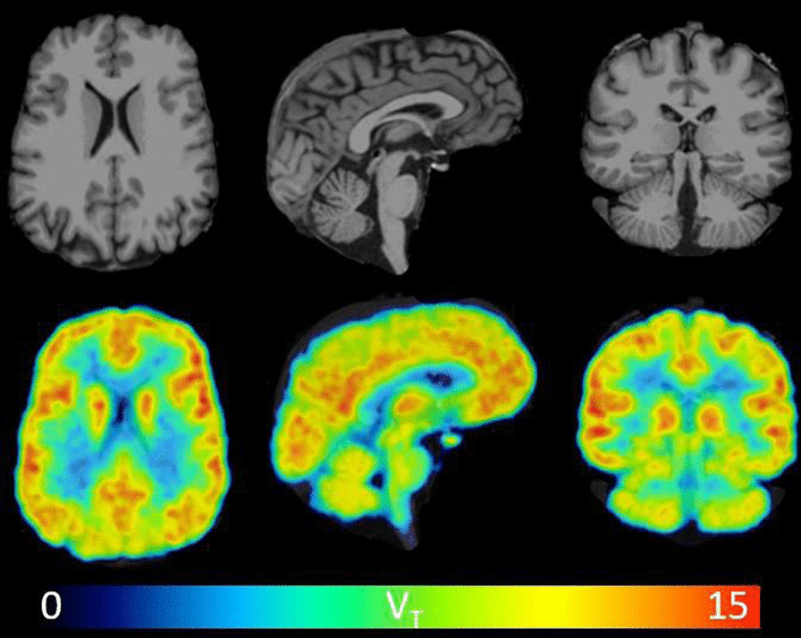
Fig. 4.

Visual inspection of 11C-CPPC total distribution volume (VT) values across the human brain reveals thalamus as one of the regions of highest binding, and relatively lower binding in cerebellar cortex. Parametric VT images and MRI images in three views are shown from a representative healthy participant. VT was estimated from 90-min data using Logan graphical analysis with the metabolite-corrected arterial input function. VT is in units of mL cm−3. The VT image was filtered with a 2 mm FWHM 3D Gaussian to reduce noise
