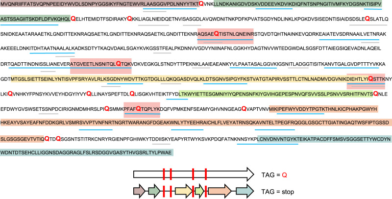
Fig. 2. Protein sequence coverage map of alternative code phage tail-related protein.
Highlighted sections of the code 15 predicted protein sequence (top) show the corresponding proteins that would have been predicted using standard code 11 (predicted open reading frames), also depicted in the graphical representation (bottom). Blue lines illustrate regions covered by tryptic peptides identified through LC-MS/MS database matching, whereas gray lines represent regions of the predicted protein sequence with matching de novo sequence tag coverage. Red text in the sequence indicates the location of glutamine residues from reassigned stop codons. Red boxes on the sequence coverage map and red bars on graphical representation indicate the recoded glutamine residues with peptides identified through database searching.

