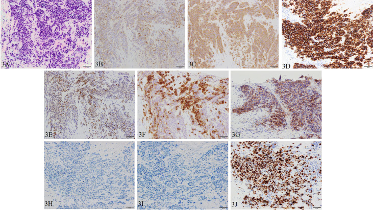
Figure 3.
Pathological manifestations of prostate re-biopsy. Hematoxylin and eosin staining (magnification, ×200): (A) the tumor tissue is infiltrated with chromatin-rich, naked nucleated tumor cells with an increased nucleus/cytoplasm ratio of irregular nuclear shape. Immunohistochemistry (magnification, ×200): (B) chromogranin A, (C) synaptophysin, (D) CD56, (E) INSM1, (F) SSTR2, and (G) PD-L1 (focal) are positive, and (H) AR and (I) PSA are negative. (J) Ki-67 is also positive with a Ki-67 labeling index over 90%.

