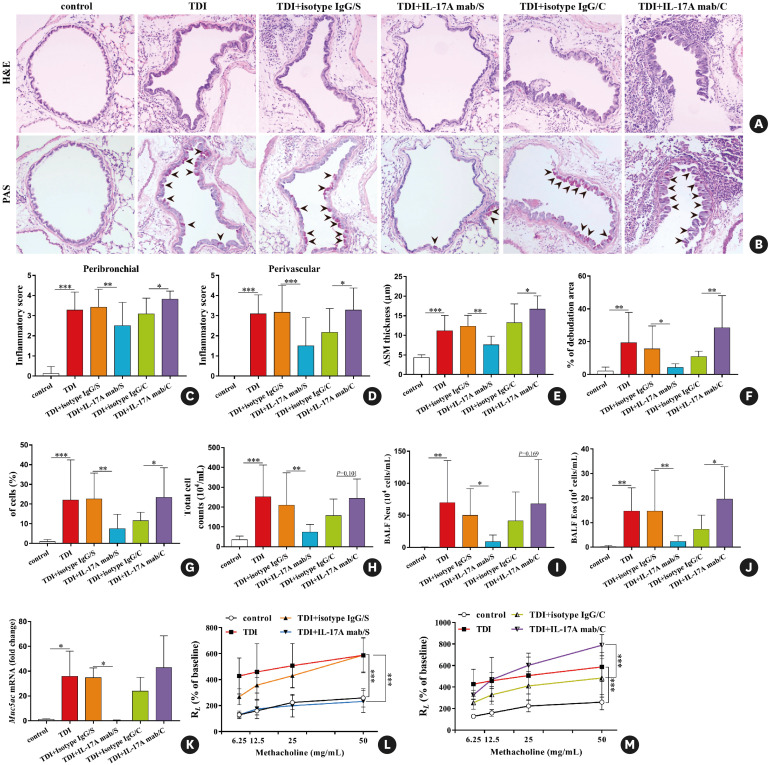
Fig. 3. Neutralization of IL-17A during antigen immunization and antigen challenge exerts distinct effects on TDI-elicited airway hyperreactivity and inflammation. (A, B) Representative H&E- and PAS-stained lung sections of different groups. Original magnification was 200×. (C, D) Semi-quantification of airway inflammation was performed (n = 8–10). (E, F) Analysis of ASM thickness and epithelial denudation was performed (n = 8–10). (G) Semi-quantification of PAS staining was performed (n = 8–10). (H-J) Numbers of total inflammatory cells, neutrophils and eosinophils in BALF (n = 8–10). (K) Expression of Muc5ac gene (quantitative PCR) in the whole lung (n = 3). (L, M) Airway hyperresponsiveness was measured by lung resistance (RL). Results are shown as percentage of baseline value (n = 5).
IL, interleukin; TDI, toluene diisocyanate; H&E, haematoxylin and eosin; PAS, periodic acid-Schiff; ASM, airway smooth muscle; BALF, bronchoalveolar lavage fluid; PCR, polymerase chain reaction; Ig, immunoglobulin.
*P < 0.05; **P < 0.01; ***P < 0.001.

