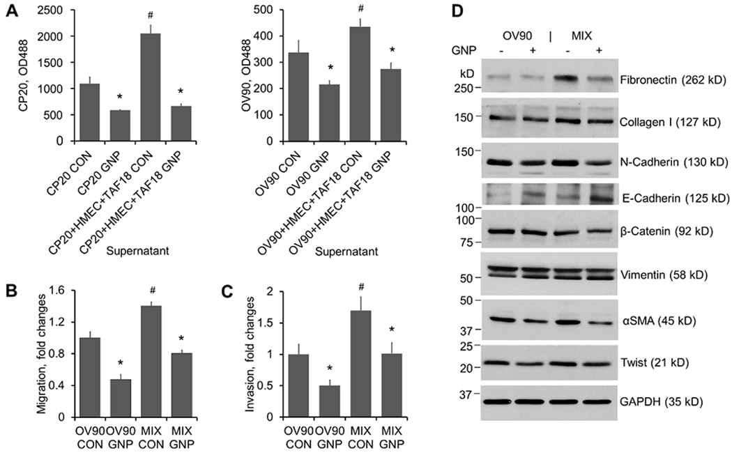
Fig 5.

Supernatants of cultures treated with GNP inhibit CC proliferation, migration, invasion and EMT. CM preparation: 5x10⁵ CP20-EGFP or OV90-EGFP cells were seeded to 10 cm dishes with or without HMEC and TAF18 of equal numbers. Cells were starved from the next day for 16 h and then treated with 25 μg/ml GNP or PBS in fresh serum-free media for 2 days. Supernatants were collected and centrifuged to remove cell debris and GNP. (A) Proliferation: CP20-EGFP or OV90-EGFP cells were seeded at 1000 cells/well to 96-well plates. The following day the CM were added to plates and incubated for 3 days. EGFP fluorescence intensity was measured after treatment. Experiments were performed in sextuplicate and repeated 3 times. (B) Migration: OV90-EGFP (designated as OV90 hereafter) cells (8x10⁴ cells) were seeded to each transwell. Migration was induced by CM added to the outwells for 16 h. Experiments were performed in duplicate and repeated 3 times. (C) Invasion: OV90 cells (1x10⁵ cells) were seeded to each Matrigel precoated transwell. Invasion was induced by CM added to the outwells for 24 h. Experiments were performed in duplicate and repeated 3 times. (D) EMT marker expression change upon CM treatment. OV90 cells were treated for 2 days with CM from OV90 alone or from the OV90, TAF18 and HMEC cocultures (MIX) incubated with or without GNP. Proteins (5 μg- 50 μg) in cell lysates were separated with 6% or 12% SDS-PAGE. GAPDH was used as loading control. Experiments were repeated 3 times. *, p<0.05, compared to corresponding control. #, p<0.05, compared to CP20 CON or OV90 CON.
