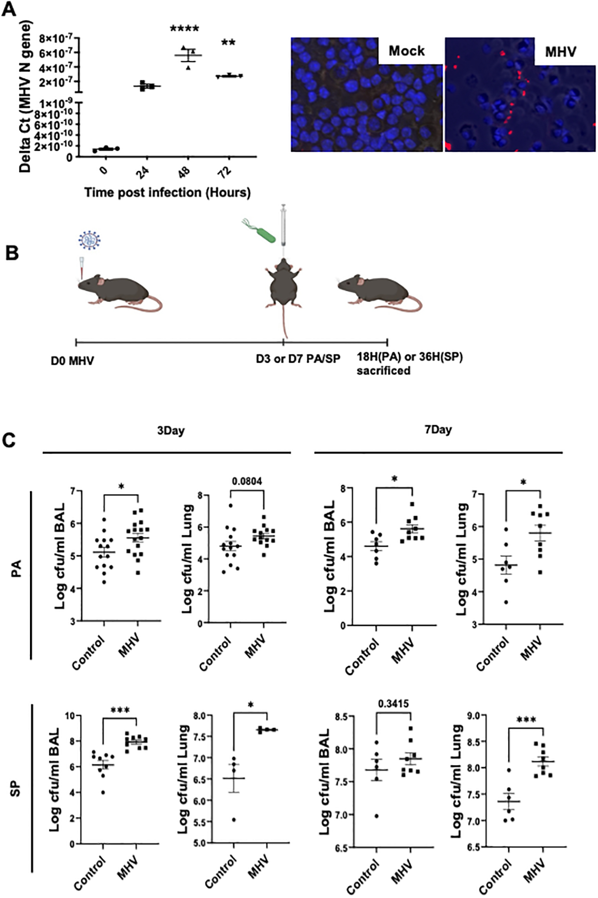
Fig. 1. MHV infects immune cells and impairs bacterial clearance in Gram-positive and Gram-negative bacterial lung infections.

(A) Mouse alveolar macrophages were obtained by lavaging naïve mice and cells were seeded at 1 × 10⁵ cells/well in 96 well plate. Cells were then infected with MHV (MOI of 10) for 2 hours and washed twice to remove the residual viruses and replenished with 300 μl of fresh media. A 50 μl aliquot was obtained at 0, 24, 48, and 72 hours and RNA was purified to run qPCR using primers for the MHV N gene. Data are from one of two independent experiments with similar outcomes and represented as Delta Ct values (A, left). To confirm that MHV infects lung immune cells in vivo, we stained the BAL cells obtained from either mock or MHV-infected mice on day 3. Representative images are shown (A, right) from two independent experiments. Red- MHV N protein, blue-DAPI. (B) Mouse model of post coronaviral bacterial infection. Mice infected with MHV (intranasal) for 3 or 7 days were superinfected with Pseudomonas aeruginosa (PA, 2.5 × 10⁶ CFUs) or Streptococcus pneumoniae (SP, 1 × 10⁴ CFUs) through the intratracheal route and then euthanized at 18 hours (PA) or 36 hours (SP) post-bacterial infection. (C) Bacterial load in the BAL and lung tissue (left lobe) was measured. Data are pooled from two or three independent experiments. *, P < 0.05, **, P < 0.01, ***, P < 0.005, ****, P < 0.0001 using either t-test or one-way ANOVA followed by Dunnett’s multiple comparisons test, as appropriate.
