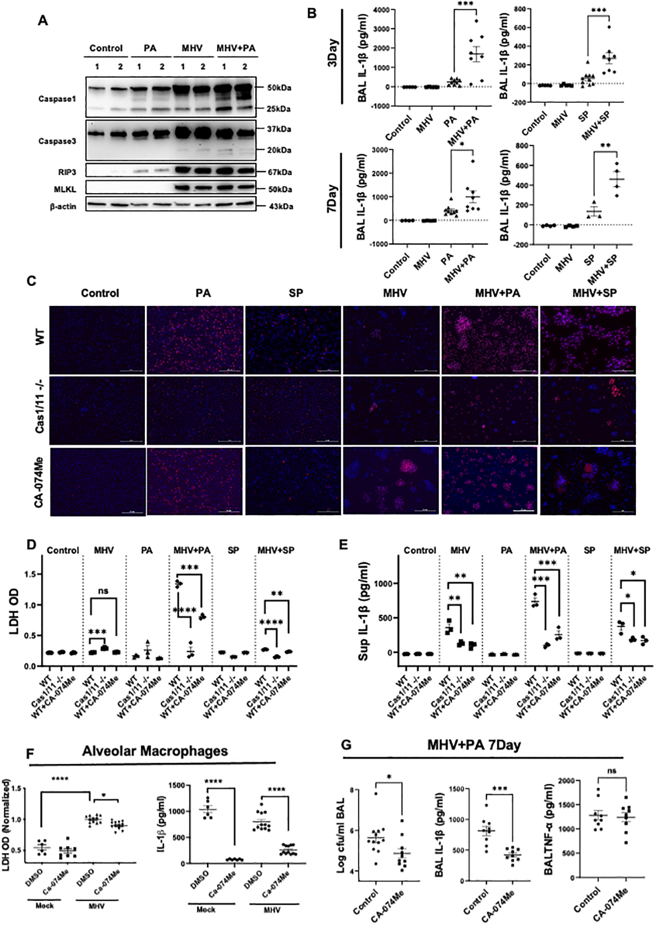
Fig. 4. Pyroptosis is a key pathological cell death mechanism potentiated by MHV in presence of bacterial superinfection.

(A) Western Blot analysis to detect markers of cell death pathways in the lung tissue, including pyroptosis (Caspase-1), apoptosis (caspase-3), and necroptosis (RIP3 and MLKL) activation in lung lysates during PA infection post-MHV infection. (B) Levels of IL-1β in BAL samples from mice with bacterial infection with or without MHV. Data are pooled from two experiments. (C) Representative pictures of PI staining showing cell death in wild-type peritoneal macrophages treated with or without CA-074Me and caspase1/11−/− peritoneal macrophages in response to MHV+PA. MHV-infected cells appear to be larger given the formation of syncytia. Experiments were repeated 3 times and data from a representative experiment is shown. Scale bars are 100 μm. (D & E) The quantification of LDH and IL1-β in cell supernatants from (C). (F) LDH and IL-1β levels were also measured in the alveolar macrophages obtained from either mock or MHV infected mice that were treated with either vehicle or CA-074Me 30 minutes prior to PA infection (MOI of 10). (G) Bacterial load, IL-1β, and TNF-α in BAL from superinfected mice with the treatment of CA-074Me or vehicle were measured. Data are pooled from two independent experiments. PI = red and blue = nuclei. *, P <0.05, **, P < 0.01 ***, P <0.001 using either t-test or one-way ANOVA followed by Dunnett’s multiple comparisons test, as appropriate.
