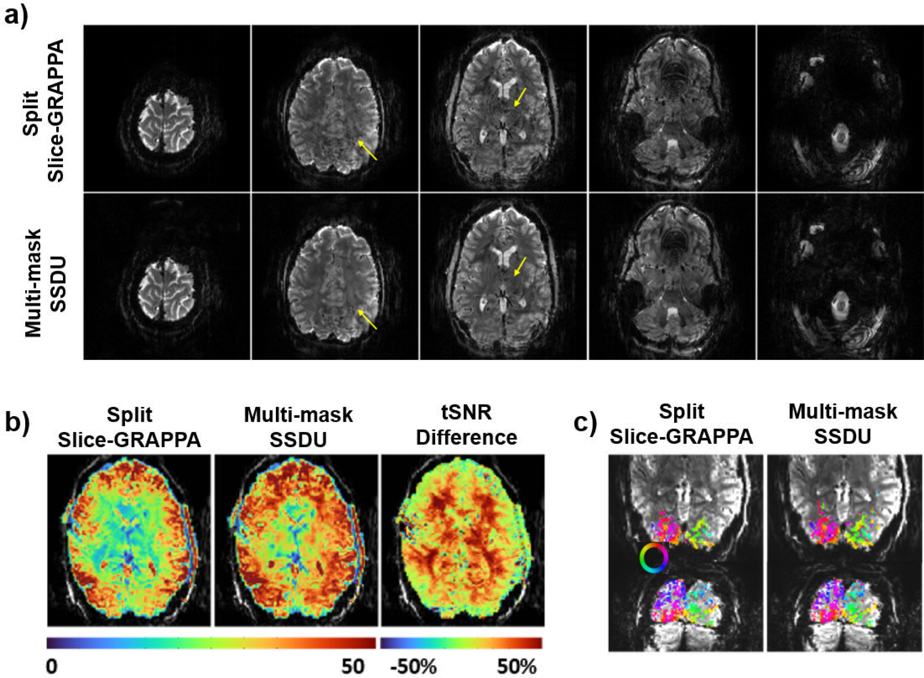
Fig. 4.

Reconstruction results from an fMRI application [6] using conventional split-slice GRAPPA technique and self-supervised multi-mask SSDU method [14]. (a) Split-slice GRAPPA exhibits residual artifacts in mid-brain (yellow arrows). Multi-mask SSDU alleviates these, along with visible noise reduction. (b) Temporal SNR (tSNR) maps show substantial gain with the self-supervised deep learning approach, particularly for subcortical areas and cortex further from the receiver coils. (c) Phase maps for the two reconstructions show strong agreement, with multi-mask SSDU containing more voxels above the coherence threshold.
