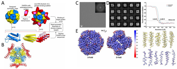
Figure 38.
(A) Schematic diagram for a scaffolding system built on a designed symmetric protein cage. (B) Cryo-EM structure of DARP14 symmetric cage core with subunits colored as in A (PDB ID: 6C9K). (C) Representative motion-corrected micrographs of DARP14. (Inset) Fourier transformation showing visible thon rings to ∼3 Å. (D) Reference-free 2D class averages highlighting good alignment of the cage and clear density for fused 17 kDa DARPins. (E) Overview of a ∼3.1 Å reconstruction of the cage core. Left: representations of unfiltered local resolution viewing down the 3-fold and 2-fold symmetry axis highlighting extensive areas at an atomic resolution of ∼2.5 Å. Top right: Fourier shell correlation curves of unmasked and masked reconstructions. Bottom right: refined models fit into density for subunit A (yellow) and subunit B (blue) of the cage core. Reprinted with permission from ref (830). Copyright 2018 National Academy of Sciences.

