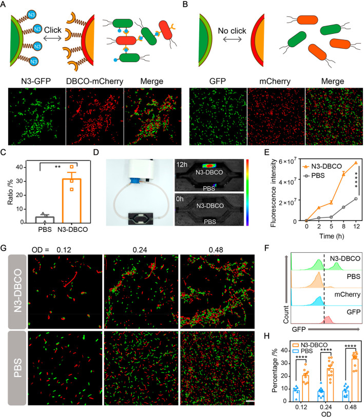
Figure 3.
In vitro characterization of bacterial aggregation induced by bioorthogonal reactions. (A) and (B) Representative confocal fluorescence images of bioorthogonal-mediated bacterial adhesion between N3-GFP + DBCO-mCherry and GFP + mCherry at a 1:1 ratio (OD = 0.48). Scale bar, 1 μm (n = 3). (C) Aggregation ratio analysis by the particle analyzing function of ImageJ. (D) In vitro bioorthogonal-mediated binding effect of N3-GFP and DBCO-mCherry in a flow environment. DBCO-mCherry was added to the circulating fluid, and the red fluorescence (mCherry) of hydrogel containing N3-GFP was monitored by IVIS (n = 3). (E) Fluorescence intensities at different time points. (F) Flow cytometry analysis for evaluating bacterial aggregation caused by bioorthogonal reactions (n = 3). (G) Representative confocal fluorescence images and (H) aggregation ratios of 1:1 mixed cocultures at different bacterial densities (n = 6). Scale bar, 1 μm. Significant differences were assessed in (C), (E), and (H) using the t test (**P ≤ 0.01; ****P ≤ 0.0001). The mean values and SEM are presented.

