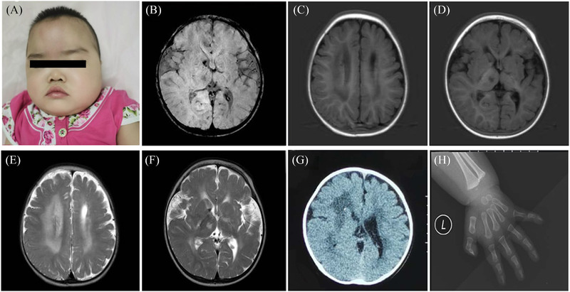
FIGURE 1.

Clinical features and images of the patient with PLOD3 mutation. (A) The patient showed hypertelorism, an upturned nose, and low‐set ears. (B–F) Cerebral magnetic resonance imaging (MRI) of the patient revealed abnormal signals in the bilateral thalamus, basal ganglia, and corona radiata, with multiple intracranial malacias and bleeding foci. White matter volume was decreased in the left cerebral hemisphere. (G) Cranial CT revealed calcification in the anterior ventricle. (H) X‐ray of the left hand showed the distal end of the metacarpal bone was irregular.
