Fig. 1.
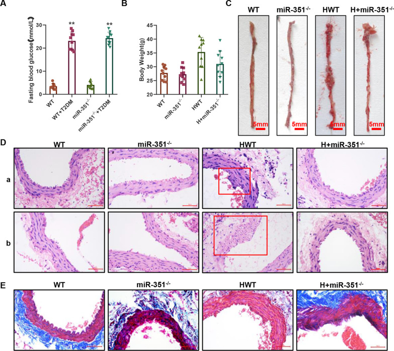
A mouse model of atherosclerosis was established, and pathological examination was conducted. A One-touch detection of the fasting blood glucose FBG index in mice. N = 10 mice per group. B Weight results of mice in each group. N = 10 mice per group. C Oil red O was used to stain the aorta in the thoracic region of mice in each group, and red stain indicated the site of oil presence or plaque formation. D HE staining results: The vascular tissue of the abdominal aorta is shown in the upper part, and the aortic arch is shown in the lower part. The scale bar represents 50 μm. E Masson trichromatic staining was performed on collagen, with blue representing collagen parts and red representing muscle fibers and other tissues. The scale bar represents 50 μm. Histopathological test, N = 3 mice per group

