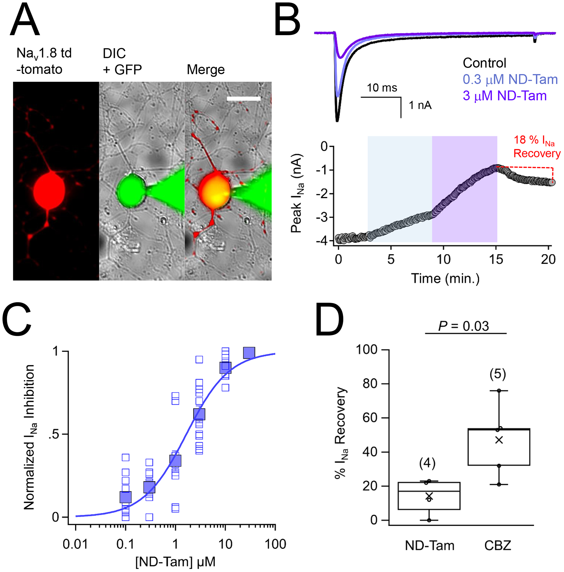
Figure 1. Tamoxifen metabolites inhibit endogenous voltage-gated sodium channels expressed in murine DRG neurons.

A) DIC and fluorescence images of voltage clamped DRG neuron isolated and cultured from an NaV1.8-tomato mouse. Continuity with the patched neuron is indicted by the GFP (Alexa Fluor 488 dye 1 nM) loaded into the glass patch electrode. Scale bar = 25 μm B) Top, exemplar sodium currents (INa) recorded in control saline and two concentrations of N-desmethyl tamoxifen (ND-Tam) from a voltage-clamped DRG neuron. INa was activated by 0.2 Hz train of 50 ms depolarizations to −10 mV from −100 mV holding potential. Bottom, time course of peak INa inhibition during control conditions and after 5 minutes of extracellular ND-Tam treatment. C) Resulting drug concentration- INa inhibition relationship for ND-Tam fit to the Hill equation. Open symbols represent responses from individual cells and filled symbols represent average response per concentration. Error is equal to S.E.M. (n = 11) D) A comparison of the percent INa recovery after 3–10 μM ND-Tam or 1 mM carbamazepine (CBZ) inhibition. INa recovery was assessed after 5 minutes in control saline post drug exposure. Fewer INa recovered from ND-Tam exposure compared to CBZ exposure, 14.3 ± 5.4 % and 47.2 ± 9.6 %, respectively, and was statistically significant. (P = 0.03). Error is equal to S.E.M. and the number of cells evaluated per treatment group is indicated within the parentheses.
