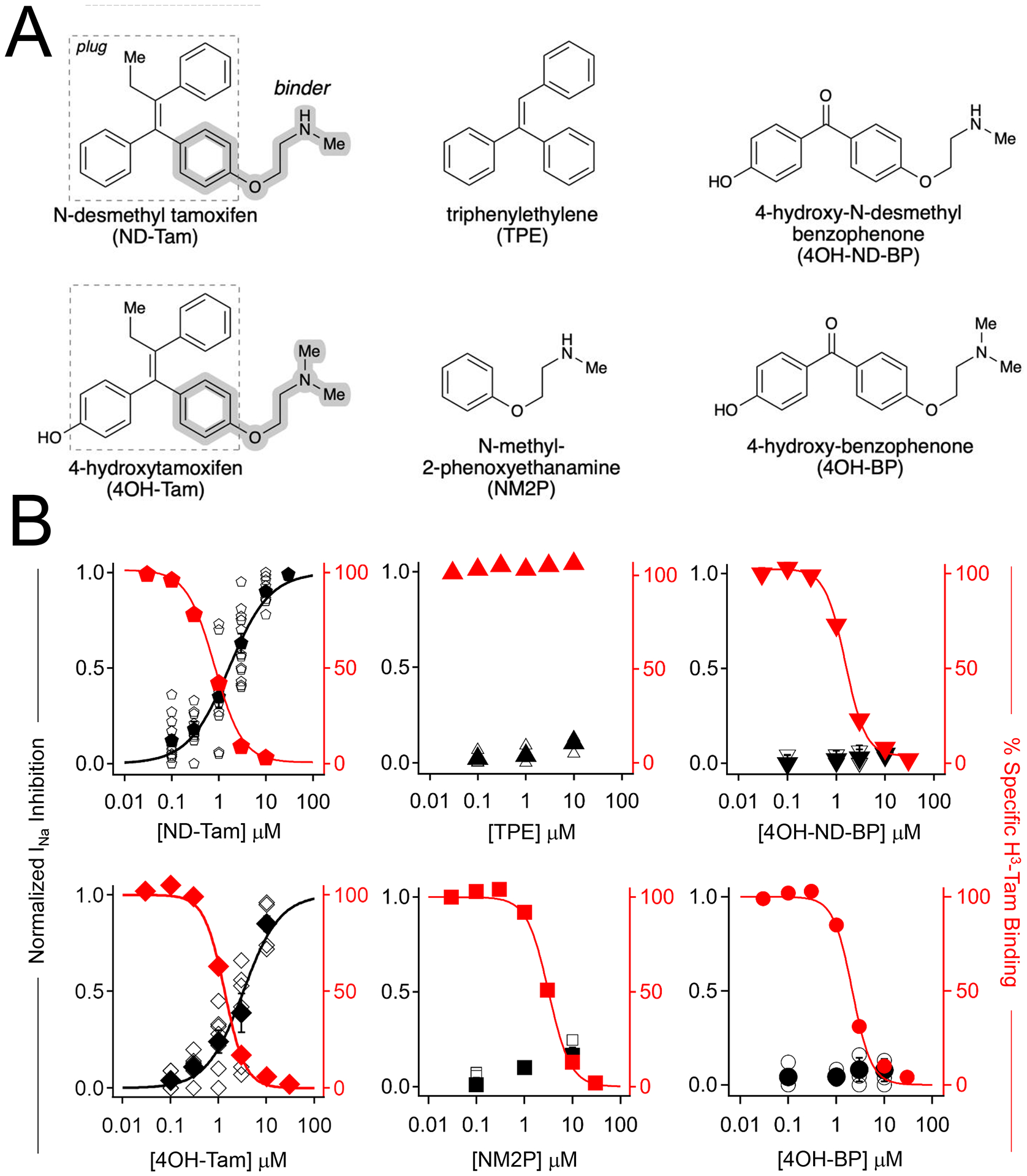
Figure 3. Structure related activity of analogs at the NaV tamoxifen receptor, as assessed by competitive binding results and DRG sodium current inhibition.

A) Molecular structures of the tested tamoxifen analogues. B) Normalized sodium current inhibition recorded DRG responses (black), and percent specific binding of tritium labeled tamoxifen (red) to membrane preparations expressing NaV1.7, plotted as a function of tamoxifen analog concentration. Open symbols are results recorded from individual neurons. Filled symbols represent the average response at each concentration. Percent specific binding was averaged from four trials. Error is equal to S.E.M.
