Figure 1.
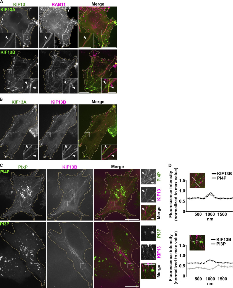
PI4P associates with KIF13+ recycling endosomal tubules. (A) Live imaging frame of a Hela cell co-expressing KIF13A-YFP and mCh-RAB11A (green and magenta, respectively; top panels) or mCh-KIF13B and GFP-RAB11A (pseudocolored in green and in magenta, respectively; bottom panels). Magnified insets (4×) show RAB11A co-distribution with KIF13+ tubules (arrowheads). (B) Live imaging frame of a Hela cell co-expressing KIF13A-YFP (green) and mCh-KIF13B (magenta). Magnified insets (4×) show KIF13A and KIF13B co-distribution (arrowheads). (C) Live imaging frame of Hela cells expressing mCh-KIF13B (magenta) together with the GFP-coupled sensors (green) for either PI4P (SidC-GFP, top) or PI3P (GFP-FYVE, bottom). Magnified insets (4×) show the co-distribution of KIF13B+ tubules with PI4P sensor (arrowheads). (D) Line scan analyses intersecting mCh-KIF13B+ tubules captured as in C (30 tubules/condition); values are mean ± SEM (n = 3 independent experiments). Cell periphery is delimited by yellow lines. Scale bars: (main panels) 10 µm; (insets) 2.5 µm.

