Figure 3.
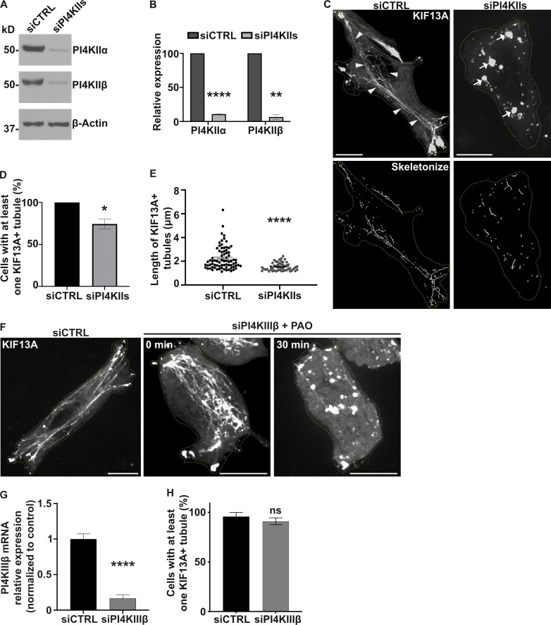
PI4KIIs are required for the formation and stabilization of recycling endosomal tubules. (A) Western blot of HeLa cell lysates treated with control (CTRL) or PI4KIIs (PI4KIIα and PI4KIIβ) siRNAs and probed for PI4KIIα (top), PI4KIIβ (middle), and β-Actin (loading control, bottom). (B) Protein expression levels of PI4KIIα and PI4KIIβ in siCTRL- or siPI4KIIs-treated cells normalized to β-Actin levels. (C) Live imaging frame (top) and associated binary images from “skeletonize” processing (bottom) of siCTRL- or siPI4KIIs-treated HeLa cells expressing KIF13A-YFP. Arrowheads, KIF13A+ RE tubules in siCTRL cells. Arrows, KIF13A+ vesicular structures in PI4KIIs-depleted cells. (D) Quantification of the average percentage of siCTRL or siPI4KIIs cells (n > 60) with at least one KIF13A-YFP+ tubule. (E) Quantification of the average length of KIF13A-YFP+ tubules (n > 50 cells) in siCTRL or siPI4KIIs cells. (F) Live imaging frame of KIF13A-YFP expressing siCTRL (left) or siPI4KIIIβ (middle and right) HeLa cells. Right panel shows siPI4KIIIβ-treated cells treated with PAO. (G) Quantification of the PI4KIIIβ mRNA expression levels in siCTRL or siPI4KIIIβ cells by quantitative RT-PCR analysis relative to GAPDH. (H) Quantification of the average percentage of siCTRL- or siPI4KIIIβ-treated cells (n > 60) with at least one KIF13A-YFP+ tubule. Cell periphery is delimited by yellow dashed lines. Data represent the average of at least three independent experiments and are presented as mean ± SEM. (B, D, E, G, and H): two-tailed unpaired t test; ns, not significant; *, P < 0.05; **, P < 0.01; ****, P < 0.0001. Scale bars: 10 μm. Source data are available for this figure: SourceData F3.

