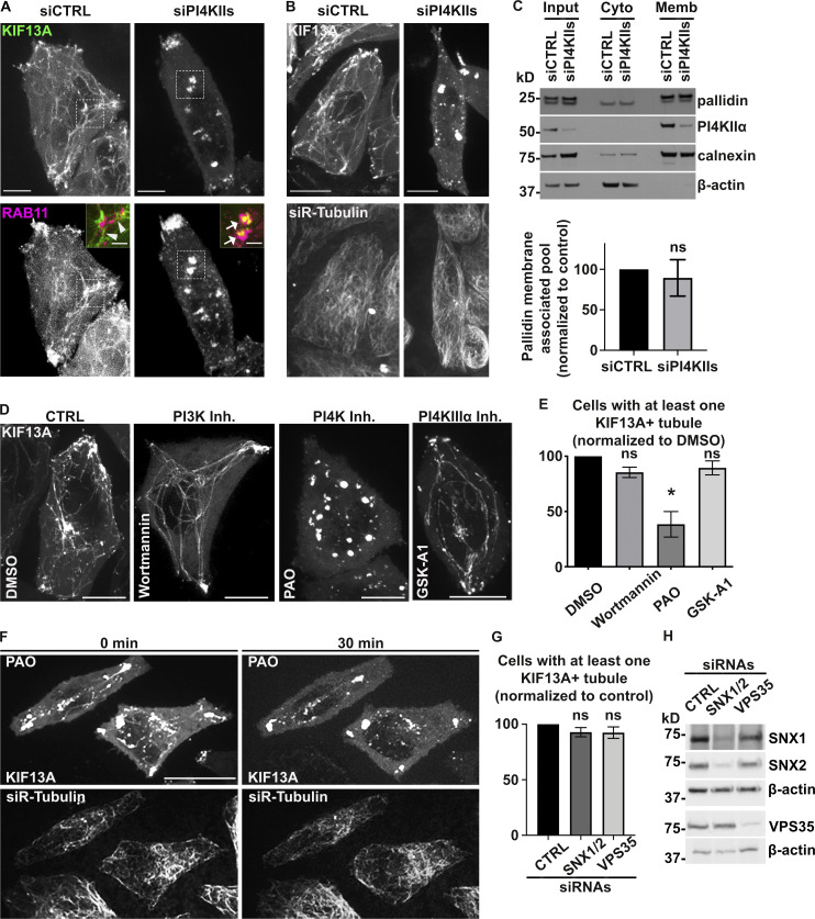
Figure S3.
PI4KIIs enzymatic activity is required for the formation and stabilization of recycling endosomal tubules. (A) Live imaging frames of siCTRL- (left) or siPI4KIIs- (right) treated HeLa cells co-expressing KIF13A-YFP (top) and mCh-RAB11A (bottom). Note the tubular (arrowheads) or vesicular (arrows) KIF13+ structures in siCTRL- or siPI4KIIs-treated cells. Insets are magnifications of boxed areas showing KIF13A and RAB11A co-distribution. (B) Live imaging frame of siCTRL- (left) or siPI4KIIs- (right) treated HeLa cells expressing KIF13A-YFP (top) and incubated with siR-Tubulin probe (bottom) to visualize microtubules. (C) siCTRL- and siPI4KIIs-treated cells were homogenized and fractionated to yield post-nuclear membrane (Memb) and cytosolic (Cyto) fractions; input before fractionation is shown at left. Identical cell equivalents of the two fractions were analyzed by immunoblotting using antibodies to membrane-associated calnexin, cytosolic β-actin, PI4KIIα, and the pallidin subunit of BLOC-1. A representative blot is shown (top). Quantification (bottom) of the percentage of membrane-associated pallidin relative to the total cellular content and normalized to siCTRL as 100 (siPI4KIIs: 89.4 ± 22.4). (D) Live imaging frame of HeLa cells expressing KIF13A-YFP and treated for up to 30 min with DMSO vehicle (first panel), PI3K inhibitor wortmannin (10 µM, second panel), PI4K inhibitor PAO (300 nM, third panel), or PI4KIIIα inhibitor GSK-A1 (100 nM, fourth panel). (E) Quantification of the average percentage of treated cells as in D with at least one KIF13A-YFP+ tubule (n > 60 cells). (F) Time lapse images of HeLa cells expressing KIF13A-YFP (top) and treated with siR-Tubulin to visualize microtubules (bottom) before (0 min) and after (30 min) PAO (600 nM) addition. (G) Quantification of the average percentage of KIF13A-YFP+ HeLa cells treated with CTRL, SNX1 and SNX2 (SNX1/2), or VPS35 siRNAs with at least one KIF13A-YFP+ tubule (n > 30 cells). (H) Lysates of siRNA-treated HeLa cells as in G immunoblotted with antibodies to SNX1, SNX2, VPS35, or β-actin (loading control). Data represent the average of at least three independent experiments and are presented as mean ± SEM. (C, E, and G) Two-tailed unpaired t test; *, P < 0.05. ns, not significant. Scale bars: (main panels) 10 µm; (magnified inset) 2.5 µm. Source data are available for this figure: SourceData FS3.

