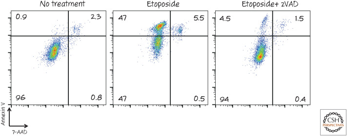
Figure 3.
Detection of phosphatidylserine exposure by fluorescence-activated cell sorting (FACS). Cells were treated with the chemotherapy agent etoposide to induce apoptosis (± the caspase inhibitor zVAD-fmk) and then stained with annexin V (coupled with a fluorescent dye) and the vital dye 7-AAD. The fluorescence intensity of each cell is represented by a dot. Notice that the cell population becomes positive for annexin V before membrane integrity (measured by 7-AAD) is lost. This annexin V staining, and by implication phosphatidylserine exposure, is dependent on caspases, as treatment with the inhibitor zVAD largely prevented it. 7-AAD, 7-amino actinomycin D; zVAD, zVAD-fmk.

