. 2022 Sep 30;106(1):88. doi: 10.5334/jbsr.2891

Figure 2.

MRI at 3T performed three weeks later, because of a second stroke-like event. A similar lesion in the gyri of the left occipital lobe is present. Signal abnormalities in the previously affected area nearly normalized, but microhemorrhages persisted

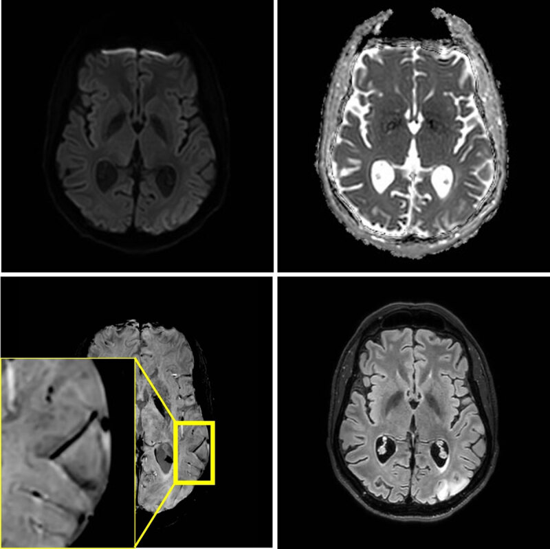
MRI at 3T performed three weeks later, because of a second stroke-like event. A similar lesion in the gyri of the left occipital lobe is present. Signal abnormalities in the previously affected area nearly normalized, but microhemorrhages persisted.