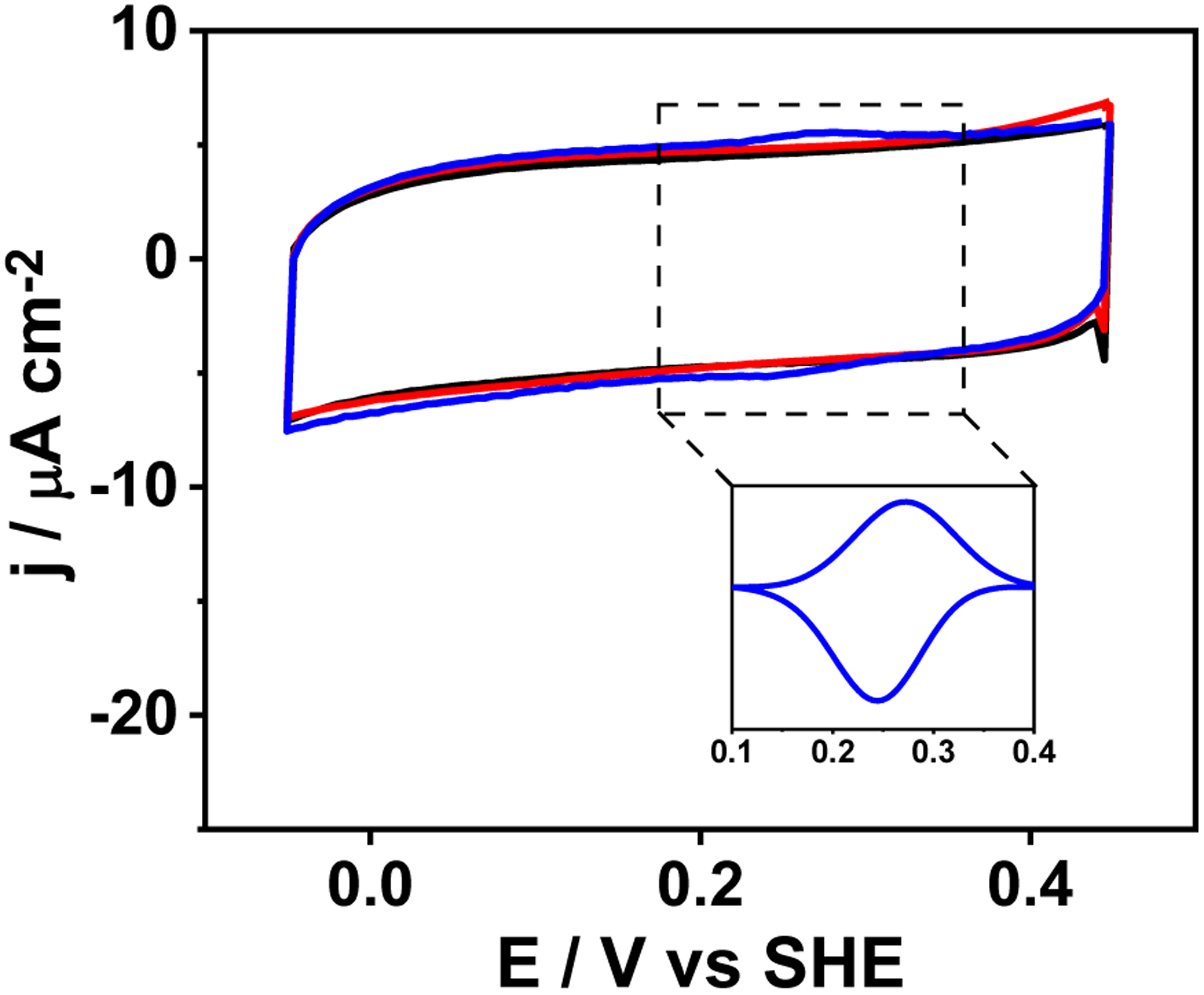
Figure 6.

PFV scans of PGE (black trace), [3SCC-(I9H)3] (red trace), [3SCC-Cu(I9H)3]2+ (blue trace) at 50 mVs−1 in N2-saturated 80 mM mixed buffer at pH 6.5. The inset shows background corrected peak for [3SCC-Cu(I9H)3]2/1+. Peptide samples were dropped on PGE and dried under N2 prior to data collection. Data were collected against Ag/AgCl reference electrode and converted to SHE.
