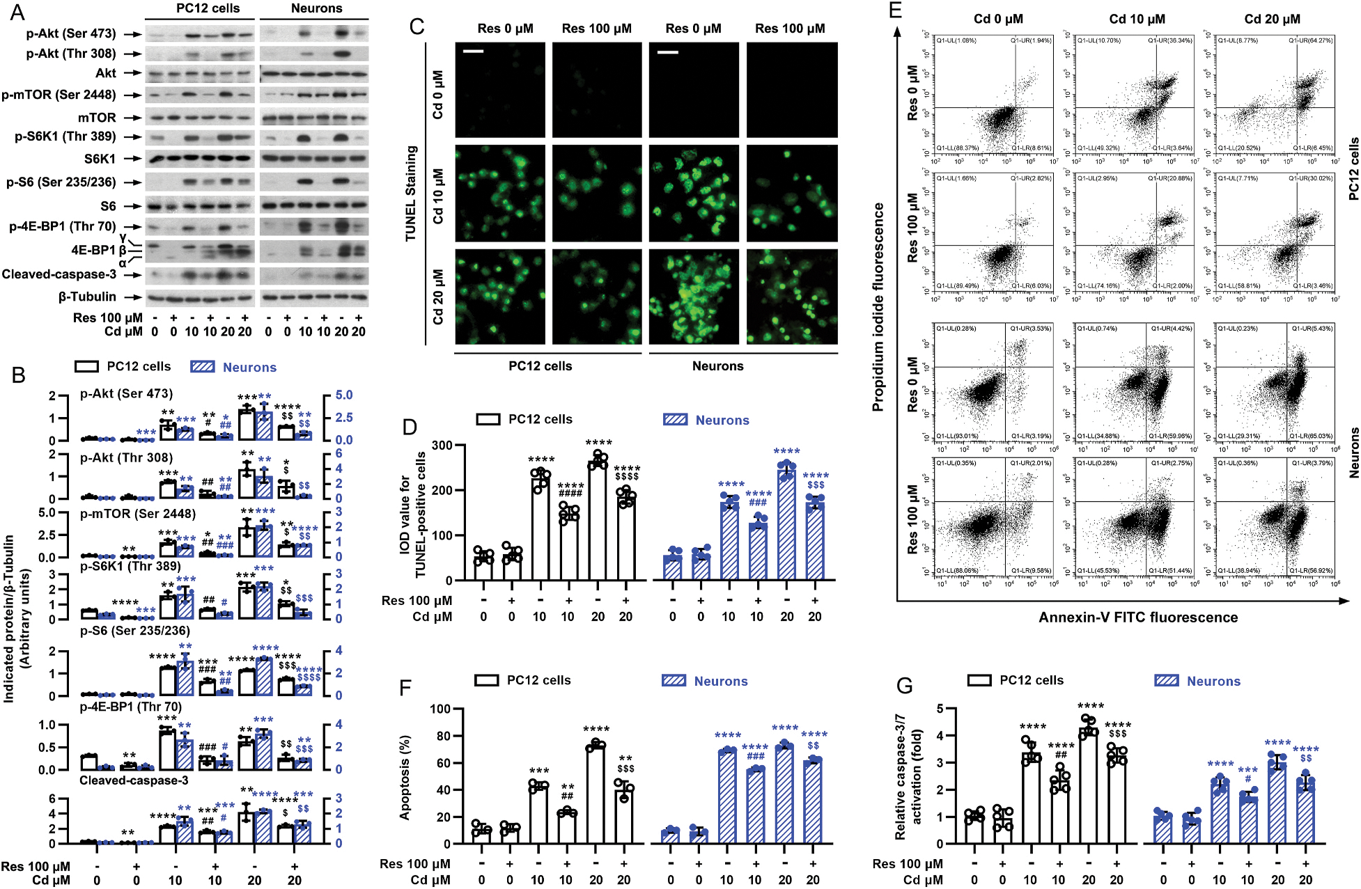
Fig. 2.

Pretreatment with resveratrol mitigates Cd-induced activation of Akt/mTOR pathway and cell apoptosis in neuronal cells. PC12 cells and primary neurons were pretreated with/without resveratrol (Res, 100 μM) for 1 h, followed by exposure to Cd (10 and 20 μM) for 4 h (for Western blotting) or 24 h (for TUNEL staining, annexin-V-FITC/PI staining and caspase-3/7 activity assay). A) Resveratrol substantially repressed Cd-induced phosphorylation of Akt, mTOR, S6K1, S6 and 4E-BP1, as well as cleavage of caspase-3 in PC12 cells and primary neurons. Total cell lysates were subjected to Western blot analysis using indicated antibodies. The blots were probed for β-tubulin as a loading control. Similar results were observed in at least five independent experiments. B) The relative densities for p-Akt (Ser473), p-Akt (Thr308), p-mTOR (Ser2448), p-S6K1 (Thr389), p-S6 (Ser235/236), p-4E-BP1 (Thr70), cleaved-caspase-3 to β-tubulin were semi-quantified using NIH image J. C) Apoptotic cells were evaluated by in situ detection of fragmented DNA (in green) using TUNEL staining. Scale bar: 20 μm. D) IOD values of TUNEL-positive cells with the fluorescence staining were quantified, showing that resveratrol markedly attenuated Cd-induced apoptosis in PC12 cells and primary neurons. E) The percentages of necrotic (Q1-UL), late apoptotic (Q1-UR), live (Q1-LL) and early apoptotic (Q1-LR) cells were determined by FACS using annexin-V-FITC/PI staining. The results from a representative experiment in PC12 cells are shown. F) Quantitative analysis of apoptotic cells by FACS assay. G) Caspase-3/7 activity was determined using Caspase-3/7 Assay Kit, showing that resveratrol significantly blocked Cd-activation of caspase-3/7 in the cells. Results are presented as mean ± SEM, n = 3–5. *p < 0.05, **p < 0.01, ***p < 0.001, ****p < 0.0001, difference vs control group; #p < 0.05, ##p < 0.01, ###p < 0.001, ####p < 0.0001, difference vs 10 μM Cd group; $p < 0.05, $$p < 0.01, $$$p < 0.001, $$$$p < 0.0001, difference vs 20 μM Cd group.
