
Fig. 8.
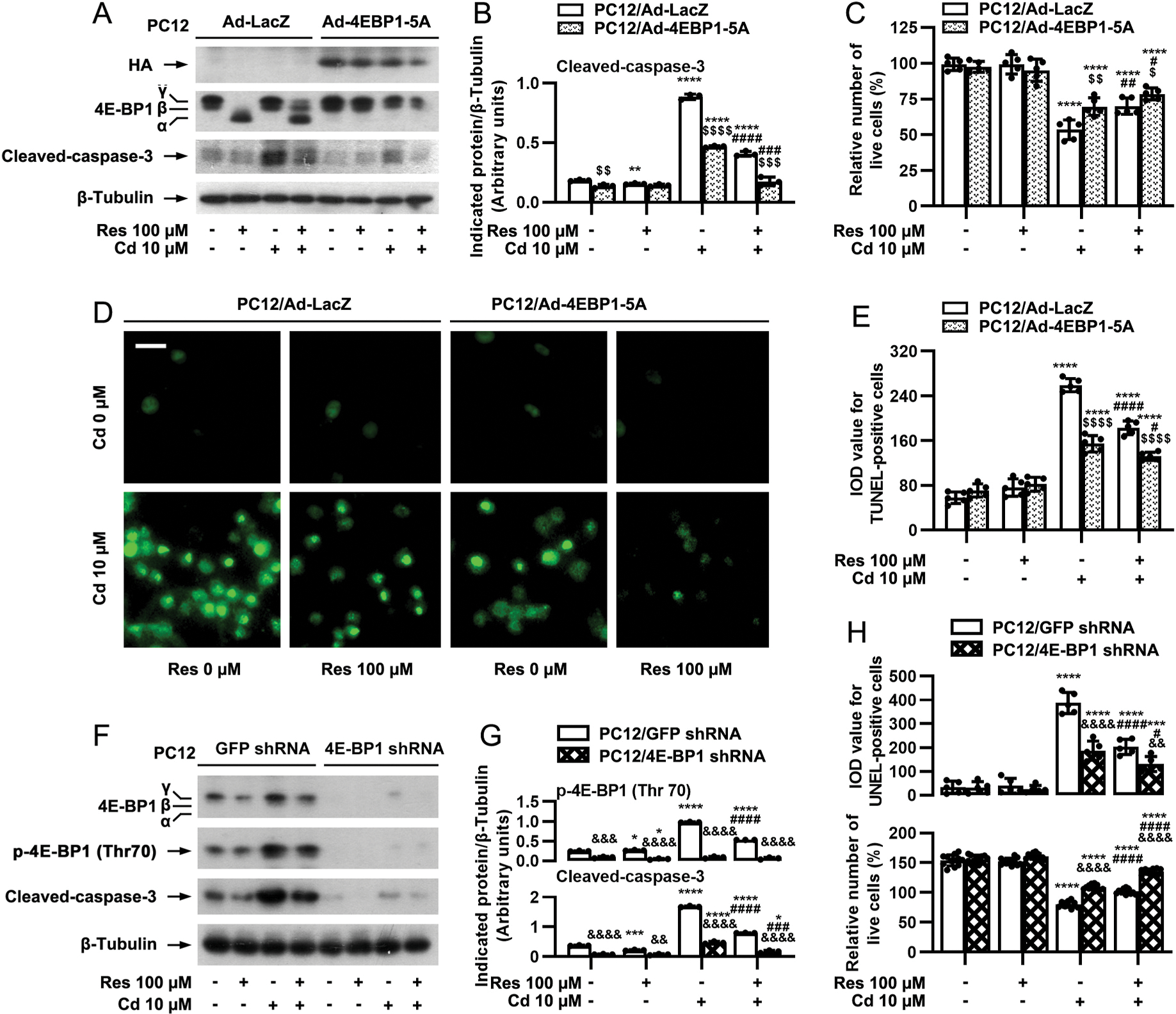
mTORC1-mediated 4E-BP1 pathway participates in resveratrol’s rescue from Cd-induced apoptosis in neuronal cells. PC12 cells, infected with Ad-4EBP1–5A or Ad-LacZ (as control), or with lentiviral shRNA to 4E-BP1 or GFP (as control), were pretreated with/without resveratrol (Res, 100 μM) for 1 h, followed by exposure to Cd (10 μM) for 4 h (for Western blotting) or 24 h (for live cell assay and TUNEL staining). A and F) Total cell lysates were subjected to Western blot analysis using indicated antibodies. The blots were probed for β-tubulin as a loading control. Similar results were observed in at least five independent experiments. B and G) The relative densities for p-4E-BP1 (Thr70), cleaved-caspase-3 to β-tubulin were semi-quantified using NIH image J. C, D, E and H) Live and apoptotic cells were evaluated by counting viable cells using trypan blue exclusion and by in situ detection of fragmented DNA using TUNEL staining, respectively. Results are presented as mean ± SEM, n = 3–5. *p < 0.05, **p < 0.01, ***p < 0.001, ****p < 0.0001, difference vs control group; #p < 0.05, ##p < 0.01, ###p < 0.001, ####p < 0.0001, difference vs 10 μM Cd group; $p < 0.05, $$p < 0.01, $$$p < 0.001, $$$$p < 0.0001, Ad-4EBP1–5A group vs Ad- LacZ group; &p < 0.05, &&p < 0.01, &&&p < 0.001, &&&&p < 0.0001, 4E-BP1 shRNA group vs GFP shRNA group.