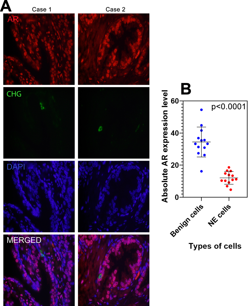
Figure 1: AR expression in benign NE cells.
(A) Representative AR and chromogranin A immunostaining in formalin fixed paraffin embedded benign prostate glands. The histologic sections were subjected to indirect immunofluorescence with anti-AR (red) and anti-chromogranin (green) antibodies to detect neuroendocrine cells and quantify AR expression. Images reduced from 630X. (B) Absolute androgen receptor (AR) expression level comparison between benign luminal and benign neuroendocrine cells. p-value by paired T-test.

