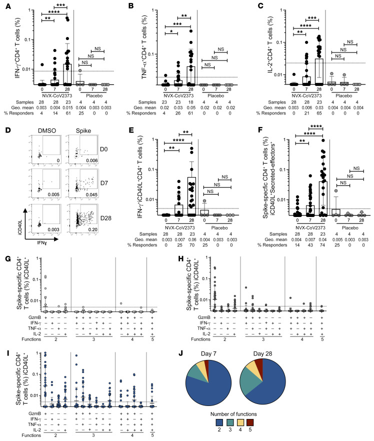
Figure 2. Cytokine-producing spike-specific CD4+ T cell responses following NVX-CoV2373 vaccination.
Proportion of (A) IFN-γ+, (B) TNF-α+, and (C) IL-2+ spike-specific CD4+ T cells detected following peptide stimulation. (D) Representative FACS plots and (E) proportion of IFN-γ+ intracellular CD40L+ (iCD40L+) responses in spike-specific CD4+ T cells on days 0, 7, and 28 after vaccination. (F) Proportion of spike-specific CD4+ T cells expressing iCD40L and producing IFN-γ, TNF-α, IL-2, or GzmB (“secreted-effector+”). Predominant multifunctional profiles of spike-specific CD4+ T cells with 1, 2, 3, 4, or 5 functions were analyzed on (G) day 0 (D0), (H) D7, and (I) D28 after vaccination. (J) Pie charts depicting the proportion of spike-specific CD4+ T cells exhibiting 2, 3, 4, or 5 functions on day 7 and day 28 after immunization. Functionality is defined as a cell expressing iCD40L and any combination of IFN-γ, TNF-α, IL-2, or GzmB. Dotted line indicates LOQ for the assay, and was calculated as the geometric mean of all sample DMSO wells multiplied by the geometric SD. Percentage responders was calculated as responses ≥ LOQ divided by the total samples in the group. Paired data were analyzed by Wilcoxon’s signed-rank test. Data shown as geometric mean ± geometric SD. *P < 0.05; **P < 0.01; ***P < 0.001; ****P < 0.0001.

