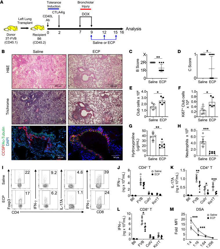
Figure 1. ECP prevents BOS and lymphocyte recognition of transplant antigens.
(A) 3T-FVB left lungs were transplanted into C57BL/6 (B6) mice and treated with CD40L Abs (POD0) and CTLA4 Ig (POD2) to establish allograft tolerance. Between POD7 and POD9, recipients ingested DOX. They received saline or ECP-treated B6 leukocytes on POD9, POD12, and POD15 and were euthanized on POD16. (B) Representative allograft H&E, trichrome, and CCSP/Ac-tubulin Ab staining. Images shown are representative of n = 10/group. Allografts scored for airway inflammation (C) (B score) and (D) the presence (designated 1) or absence (designated 0) of OB lesions (C score) (n = 10/group). Intragraft (E) total (n = 5/group) and (F) Ki67+ club cell numbers (n = 5/group) and (G) hydroxyproline content (n = 6/group). (H) Intragraft neutrophil numbers (n = 6/group). (I) Representative FACS plots of the intragraft percentage of abundance for indicated T lymphocyte lineages (n = 5/group). (J–L) T cell antigen specificity measured by IFN-γ and IL-17A production following stimulation with splenocytes isolated from B6 (syngeneic antigens), FVB (donor antigens), or B6 mice pulsed with lung self-antigens Col V and Kα1T (n = 5/group). (M) DSA (IgM) serum reactivity against FVB CD19+ cells at indicated dilutions (n = 10/group). Assay data shown for G and J–L are representative of at least 2 independent evaluations. Data are represented as mean ± SD. Two-sided Mann Whitney U test (C–H and J–M). *P < 0.05; **P < 0.01; ***P < 0.001.

