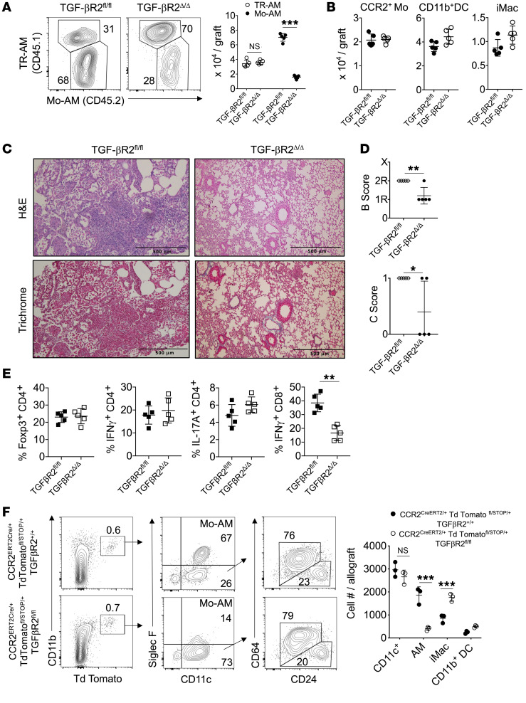
Figure 7. CCR2+ monocyte differentiation into Mo-AMs requires TGF-β leading to BOS.
TGF-βR2fl/fl and TGF-βR2Δ/Δ recipients of 3T-FVB allografts received tamoxifen i.p. every other day for 10 days, rested for 5 days, and then ingested DOX for 2 days. Eight days later, allograft recipients were analyzed for intragraft inflammation (A), as shown by representative FACS plots of the relative percentage of abundance of Mo-AMs and TR-AMs with cell counts (n = 5/group), (B) CCR2+ monocytes (Mo), CD11c+ DCs, and iMac cell counts (n = 5/group), (C) representative H&E and trichrome staining (n = 5/group), (D) airway inflammation and lesion grading (n =5/group), and (E) intragraft T cell activation (n = 5/group). (F) 3 × 10⁶ FACS-purified CCR2+ bone marrow monocytes were isolated from indicated Td Tomato reporter mice that received tamoxifen as in A and were injected into POD7 3T-FVB recipients undergoing BOS pathogenesis. On POD16, allograft tissues were quantified for Td Tomato+ Mo-AMs, CD11b+ DCs, and iMacs, as shown by representative FACS plots and cell counts. FACS plots shown are a representative result of 3 experiments. Data are represented as mean ± SD. Two-sided Mann-Whitney U test (A, B, and D–F). *P < 0.05; **P < 0.01; ***P < 0.001.

