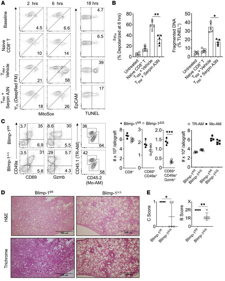
Figure 9. Gzmb+ TRM cells promote airway epithelial cell apoptosis and BOS through Blimp-1.
FVB lung epithelial cells were cocultured in a 1:2 EpCAM+ cell–to–CD8+ T cell ratio for up to 18 hours with or without Serpin A3N pretreatment (25 nM) and assessed for mitochondrial membrane potential (MitoTracker Deep Red FM), mitochondrial superoxide production (MitoSOX), and DNA fragmentation (TUNEL). Data are shown as (A) a representative FACS plot result from 5 experiments and (B) 6-hour epithelial cell mitochondrial depolarization and TUNEL activity (n = 5/condition). Blimp-1fl/fl and Blimp-1Δ/Δ recipients of 3T-FVB allografts were analyzed for intragraft inflammation as shown by (C) representative FACS plot data of TRM cell markers, Gzmb expression, and AM abundance, with cell counts n ≥ 4/group. (D) Representative H&E and trichrome staining results for n ≥ 4/group and (E) airway inflammation and lesion grading (n ≥ 4 /group). Data are represented as mean ± SD. One-way ANOVA with Dunnett’s multiple-comparison test (B); 2-sided Mann-Whitney U test (C and E).*P < 0.05; **P < 0.01.

