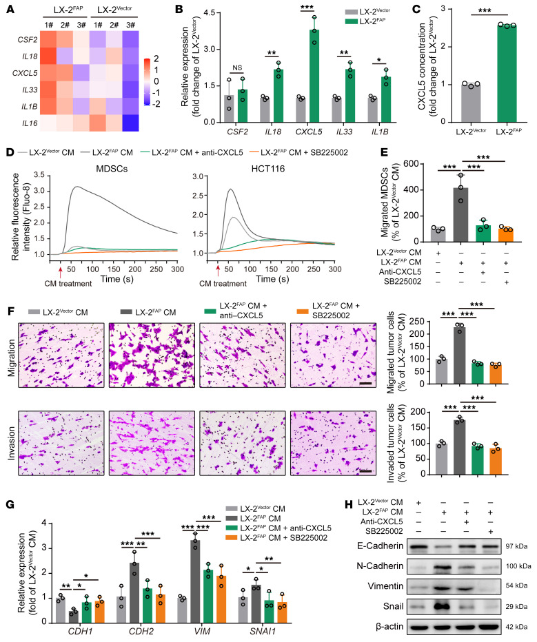
Figure 3. FAPα induces CXCL5 secretion in HSCs to promote MDSC recruitment and tumor cell EMT via activation of CXCR2.
(A) Heatmap depicting the differentially expressed genes encoding secreted factors in LX-2 cells (fold change > 1.2, P < 0.05; n = 3). (B) RT-qPCR analysis of CSF2, IL18, CXCL5, IL33, and IL1B in LX-2 cells. (C) ELISA analysis of CXCL5 in LX-2 cells (n = 3). (D) Intracellular Ca2+ mobilization in MDSCs and HCT116 cells in response to the conditioned medium from LX-2 cells in the absence or presence of CXCL5-neutralizing antibody or SB225002. (E) Transwell assay for the migration of MDSCs treated with conditioned medium from LX-2 cells in the absence or presence of CXCL5-neutralizing antibody or SB225002 (n = 3). (F) Transwell assays for the migration and invasion of HCT116 cells treated with conditioned medium from LX-2 cells in the absence or presence of CXCL5-neutralizing antibody or SB225002 (n = 3). Scale bars: 100 μm. (G) RT-qPCR analysis of CDH1, CDH2, VIM, and SNAI1 in HCT116 cells treated with conditioned medium from LX-2 cells in the absence or presence of CXCL5-neutralizing antibody or SB225002 (n = 3). (H) Western blotting analysis of E-cadherin, N-cadherin, vimentin, and snail in HCT116 cells treated with conditioned medium from LX-2 cells in the absence or presence of CXCL5-neutralizing antibody or SB225002. Data are presented as mean ± SEM. *P < 0.05; **P < 0.01; ***P < 0.001 (2-tailed, unpaired t test in B and C; 1-way ANOVA with Tukey’s post hoc comparison in E–G). CM, conditioned medium.

