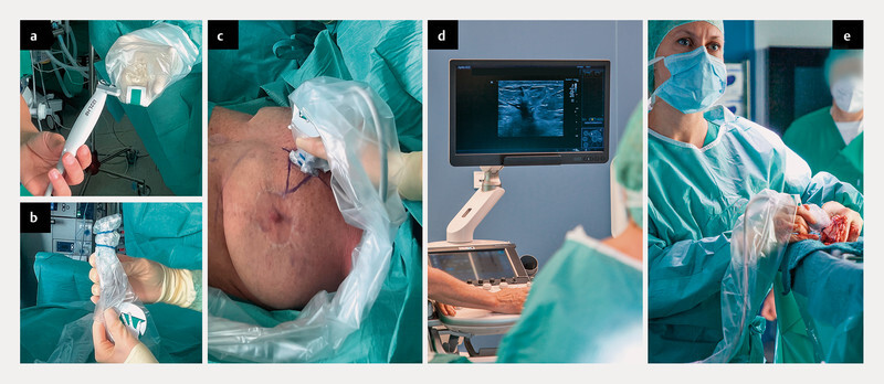
Fig. 2.

Practical use of intraoperative breast ultrasound: a The sonographic linear probe is obtained in a sterile manner. There should be sufficient gel between the probe and the film. b The sterile cover is fixed to the probe. c, d Imaging of the lesion by the surgeon. During the operation, the lesion is imaged intermittently in order to ensure a sufficient resection distance in all directions. e Immediately after removal of the tissue, the specimen is examined by ultrasound.
