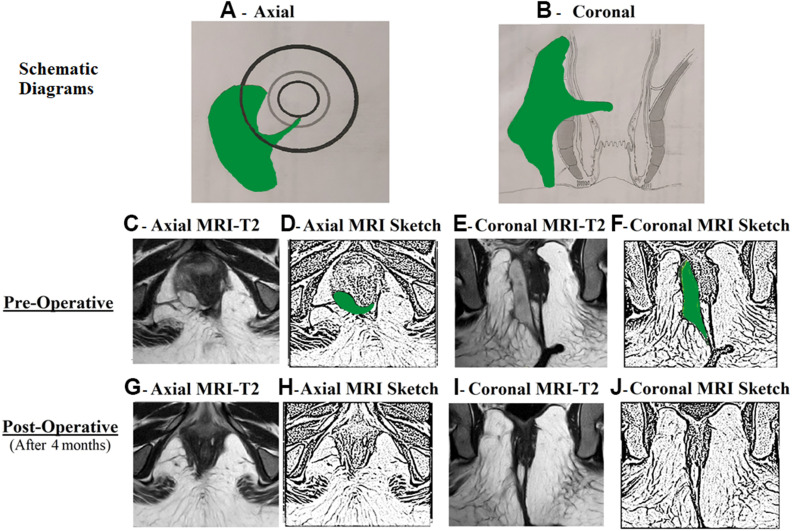
Figure 2.
A 33-year-old female patient with acute perianal abscess in right ischiorectal fossa and a high transsphincteric fistula.
Notes: The internal opening was non-locatable. MRI assessment showed that the fistula tract reached up to posterior midline (C and D). The internal opening was assumed to be at posterior midline (6 o’clock) and the fistula was managed accordingly. MRI done four months after surgery showed a completely healed fistula (G–J). (A) Axial section showing right ischiorectal abscess; (B) Coronal section; (C) Preoperative T2-weighted MRI Axial section; (D) Sketch of C; (E) Preoperative T2-weighted MRI Coronal section; (F) Sketch of E; (G) Postoperative healed T2-weighted MRI Axial section; (H) Sketch of G; (I) Postoperative healed T2-weighted MRI Coronal section; (J) Sketch of I.
Abbreviation: MRI, Magnetic resonance imaging.

