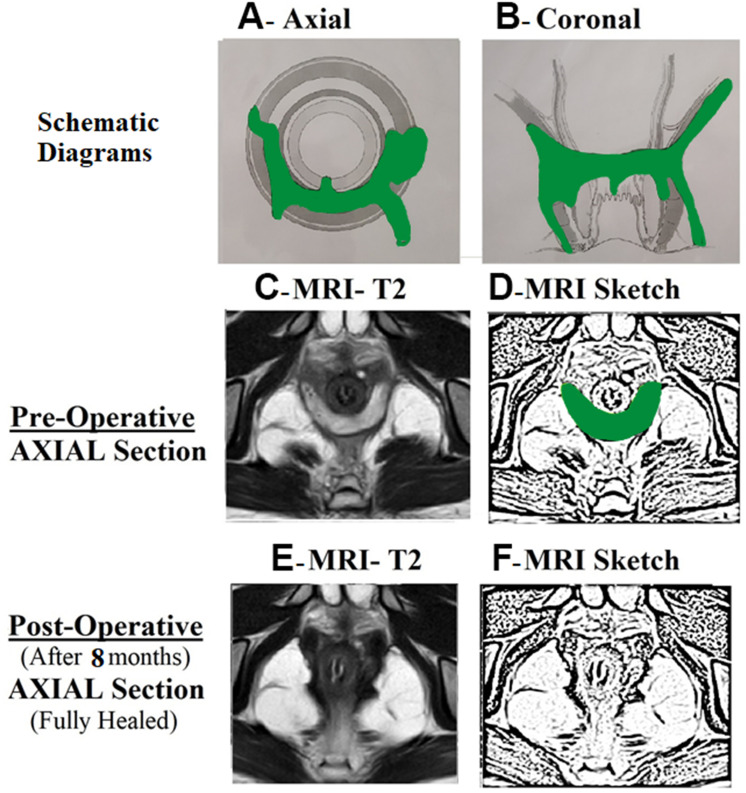
Figure 4.
A 19-year-old male patient with acute posterior horseshoe abscess.
Notes: The internal opening was non-locatable. The internal opening was assumed to be at posterior midline (6 o’clock) and the fistula was managed accordingly. MRI done eight months after surgery showed a completely healed fistula (E and F). (A) Axial section showing posterior horseshoe abscess; (B) Coronal section; (C) Preoperative T2-weighted MRI Axial section; (D) Sketch of C; (E) Postoperative healed T2-weighted MRI Axial section; (F) Sketch of E.
Abbreviation: MRI, Magnetic resonance imaging.

