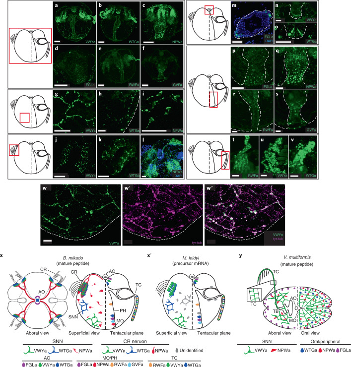
Fig. 2. Morphologies and distributions of ctenophore peptide-expressing cells.
The staining shown in each row focuses on the area enclosed by the red square in the adjacent schematic diagram. a–f, Side views of B. mikado larvae stained with antibodies against VYWa (a), WTGa (b), NPWa (c), FGLa (d), RWFa (e) and GVFa (f). g–i, High-magnification view of the SNN consisting of VWYa+ (g), WTGa+ (h) and NPWa+ (i) neurons. The dotted line in h indicates the outline of the larval body. j–l, High-magnification view of VWYa+ (j), WTGa+ (k) and NPWa+ (l) neurons at the nerve plexuses beneath comb rows. m, Aboral view of the apical organ stained by anti-FGLa antibody. The dotted line indicates the outline of the apical organ. n,o, VWYa+ (n) and WTGa+ (o) cells at the epithelial floor of the apical organ. The dotted line indicates the outline of the epithelial floor. p–s, Distribution of FGLa+ (p) NPWa+ (q), RWFa+ (r) and GVFa+ (s) cells in the pharynx. t–v, Expression of RWFa (t), VWYa (u) and WTGa (v) in the tentacle. w–w′′, Staining of VWYa+ neural network (green, w) and tyrosinated tubulin (tyr-tub; magenta, w′) and costaining of both (w′′). x,x′, Schematic diagram of the spatial distribution of amidated-peptide-expressing neurons and cells in B. mikado larva (x) and precursor-mRNA-positive neurons and cells in M. leidyi (x′). The left and right sides of the side view show a superficial view and the tentacular plane, respectively. In x, the aboral view is also shown on the left. y, Schematic diagram of the spatial distribution of amidated-peptide-expressing neurons and cells in V. multiformis adults. The left and right show the aboral side and oral side, respectively. Neurons with neurites are represented by circles with lines, and neurosecretory-like cells are represented by ovals. AO, apical organ; CR, comb row; MO, mouth; PH, pharynx; TB, tentacle bulb; TC, tentacle. Scale bar, 50 µm (a–f) or 20 µm (others). Pseudo-colours in a–w were applied using ImageJ software.

