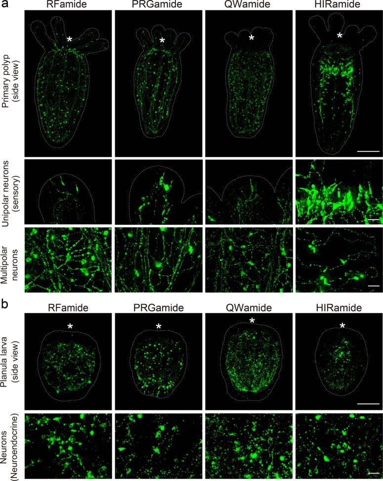
Extended Data Fig. 3. Immunostaining of neuropeptides in N. vectensis.
Expression patterns and subcellular localizations of RFa, PRGa, QWa, and HIRa of N. vectensis. a, Upper panels are side views of juvenile polyps stained by neuropeptide antibodies. Middle and lower panels show the neuronal morphologies and subcellular distributions of neuropeptides at higher magnification. Unipolar neurons with sensory cilia are located at the tentacle tip (RFa, PRGa, QWa) or the pharyngeal endomesoderm (HIRa). Multipolar neurons form subepithelial neural network (SNN) at the body column. b, Side views of late planula larvae (oral side: top). Neuropeptide-expressing neurons become visible from the larval stages. The neurons store neuropeptide-containing vesicle mainly at their cell bodies, or they do not have functional neurites, suggesting their neuroendocrine cell-like functions. Asterisks indicate oral positions. Large and small scale bars are 100 μm and 10 μm, respectively.

