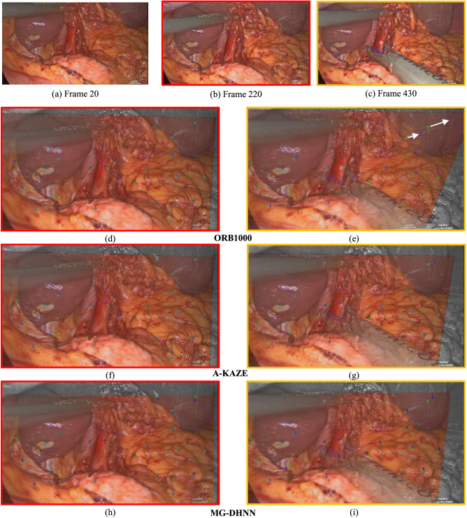
Figure 4.
Image registration results with the single homography method. (a) Frame 20 is used as the start frame of scene 1. (b) Frame 220 with small perspective changes, instrument movement, and organ deformation in relation to frame 20. (c) Frame 430 shows occlusions from an instrument and organ deformation due to manipulation compared with frame 20. (d)–(i) Semitransparent overlay of the transformed start frame 20 and the current frames 220 (left column, red) and 430 (right column, yellow) for ORB1000 (d, e), A-KAZE (f, g), and MG-DHNN (h, i). Green crosses indicate the ground truth and blue crosses the estimated position of the annotated points. White arrows highlight large registration errors and non-overlapping regions are shown in greyscale. The normalized RE averaged over the annotations of the shown frame are 0.1 for d, f, and g; 0.26 for e; 0.13 for h and i.

