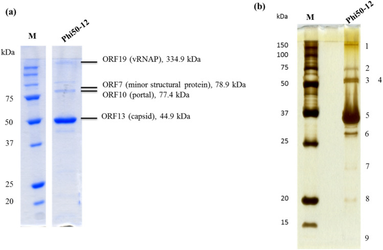
Figure 5.
Structural protein analysis by 12% SDS-PAGE. (a) Phage particles (5 × 10¹¹ PFU) were boiled in protein sample buffer (80 mM Tris, pH 6.8; 2% SDS; 2-mecraptoethanol; 0.0006% Bromophenol blue) and subjected to SDS-PAGE. Parallelly, the sliced bands were subjected to LC/MS/MS analysis. Peptides matched with the annotated ORFs, and their observed molecular mass were indicated. Four of them were identified to be virion-encapsuled RNA polymerase (ORF19), minor structural protein (ORF7), portal protein (ORF10) and capsid protein (ORF13), respectively. Original gel is presented in Supplementary Fig. S9. (b) For better visualization, a silver stain procedure was also performed, nine bands were visible on the gel. Numbers indicate the protein band location. M is the molecular weight marker, phi50-12 represents the structural protein lysate.

