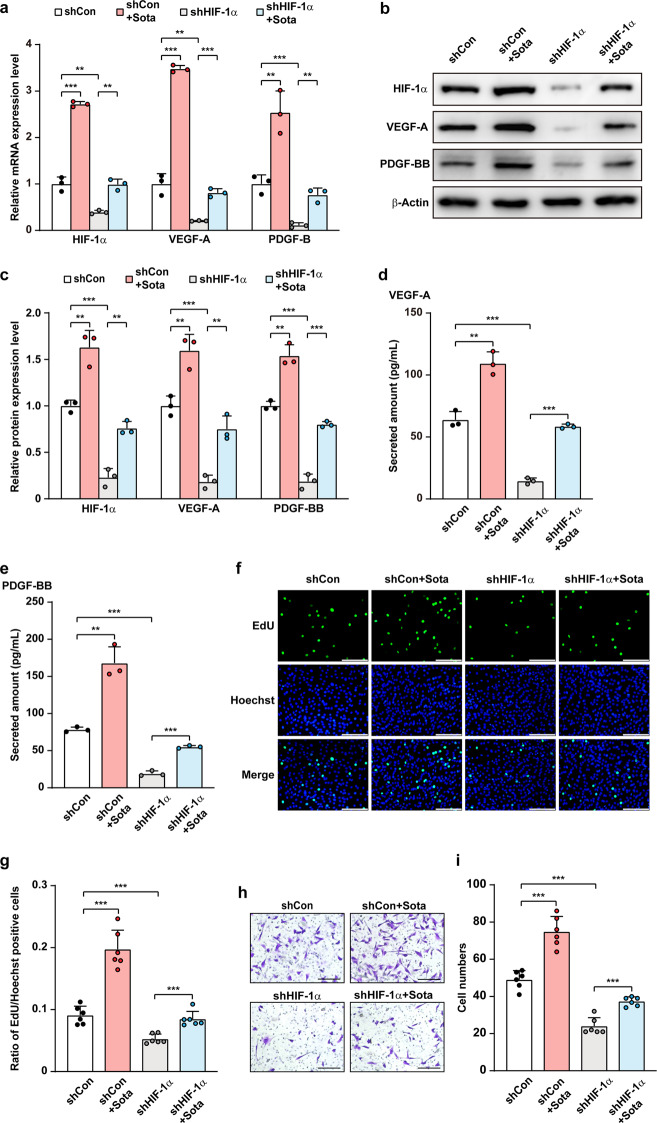
Fig. 5. Sotagliflozin promotes skeletal muscle cells angiogenesis potentials through HIF-1α under hyperglycemia.
a mRNA expression levels of angiogenic factors in HIF-1α-knocked down C2C12 cells treated with 10 μM sotagliflozin under hyperglycemia, as examined using qRT-PCR. b, c Protein expression levels of angiogenic factors in HIF-1α-silenced C2C12 cells treated with 10 μM sotagliflozin under hyperglycemia, as examined using Western blotting. Representative images (b) and quantification results (c) were shown. Secreted amount of VEGF-A (d) and PDGF-BB (e) in the culture medium of HIF-1α-knocked down C2C12 cells treated with 10 μM sotagliflozin under hyperglycemia, as analyzed using ELISA. f, g Proliferation potential of HIF-1α-knocked down C2C12 cell treated with 10 μM sotagliflozin under hyperglycemia, as evaluated by EdU incorporation assay. Representative images (f; scale bars: 100 μm) and quantification results (g; n = 6) were shown. h, i Migration potential of HIF-1α-silenced C2C12 cells treated with 10 μM sotagliflozin under hyperglycemia, as investigated using transwell migration assay. Representative images (h; scale bars: 200 μm) and quantification results (i; n = 6) were shown. Cells transfected with shCon and treated with 10% DMSO under hyperglycemia were used as controls. β-Actin was used for qRT-PCR normalization and as Western blotting loading control. Data were presented as mean ± SD (n = 3, unless further indicated). **P < 0.01; ***P < 0.001.

