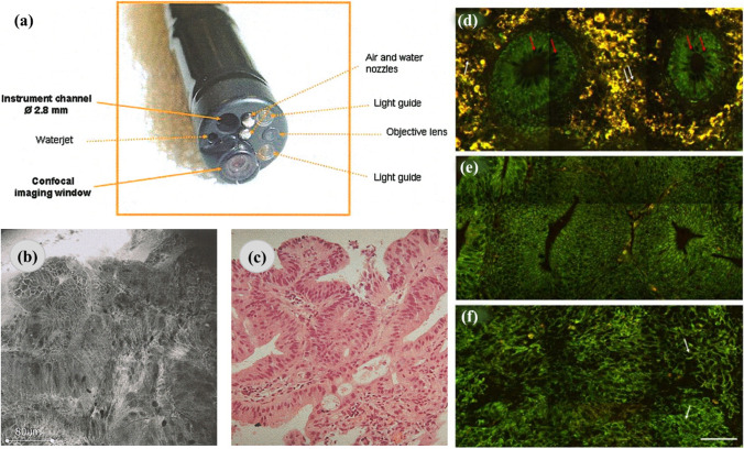
Fig. 2.
The application of confocal and multiphoton microscopy in cancer diagnosis. a A distal end of the confocal endomicroscopy. b, c Irregular cell architecture with a total loss of goblet cells and corresponding histologic specimen, respectively. Representative multiphoton images from normal (d), precancerous (e), and cancerous (f) colonic tissues at a depth of 0 μm. The excitation wavelength, λex, was 800 nm. Scale bar = 50 μm. The figures are reproduced with the kind permission from [70, 73]

