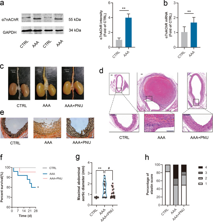
Fig. 2. Activating α7nAChR slowed down AAA formation.
Male ApoE−/− mice were infused with Ang II to induce AAA. PNU-282987 (PNU) was injected to activate α7nAChR. a, b Both protein and mRNA levels of α7nAChR were increased in aortas from AAA mice. n = 5–7 mice per group, data were shown as means ± SD, **P < 0.01 vs. CTRL. c Representative images of abdominal aortas in ApoE−/− mice, scale bar = 5 mm. d Representative images of HE staining in ApoE−/− mice, scale bar = 100 µm (up) or 50 µm (down). e Representative images of EVG staining in ApoE−/− mice. scale bar = 50 µm. f The survival curve in ApoE−/− mice. n = 20 mice for CTRL group; n = 26 mice for AAA group; and n = 22 mice for AAA + PNU group. g PNU treatment reduced maximal abdominal aortic diameters with Ang II infusion in ApoE−/− mice. n = 19–21 mice per group, data were shown as each value of per mouse. **P < 0.01 vs. CTRL; #P < 0.05 vs. AAA. h PNU treatment improved the elastin integrity, n = 12 mice per group

