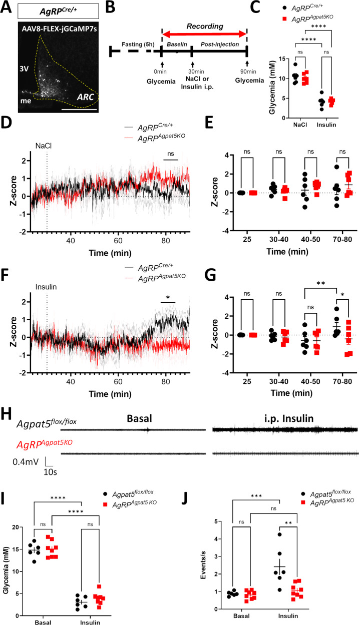
Fig. 4. Agpat5 inactivation blunts hypoglycemia-induced AgRP neuron activity and vagal nerve firing.
A Expression of GCaMP7 in the ARC of AgRPCre/+ mice following the stereotactic injection of AAV-FLEX-GCaMP7. Scale bar, 200 μm. B Scheme of the fiber photometry experiment. C Glycemia following NaCl or insulin injection in AgRPCre/+ and AgRPAgpat5KO mice, n = 6. Data are mean ± SEM, ****p < 0.0001, two-way ANOVA with Tukey’s multiple comparisons correction. D Recordings of the GCaMP7 signal in AgRP neurons of AgRPCre/+ and AgRPAgpatKO mice following NaCl injection. Data are mean ± SEM. E Quantification over 10 min periods of the GCaMP7 signal of (D). Data are mean ± SEM, two-way ANOVA with Sidak’s multiple comparisons correction. F Recordings of the GCaMP7 signal following insulin injection. Data are mean ± SEM. G Quantification over 10 min periods of the GCaMP7 signal of (F). Data are mean ± SEM, *p < 0.05, two-way ANOVA with Sidak’s multiple comparisons correction. Data are from three independent experiments, n = 6 mice. H Representative traces of parasympathetic nerve firing rate in Agpat5flox/flox (top) and AgRPAgpat5KO (bottom) mice in the basal state (left) and following insulin injection (right). I Glycemic levels of Agpat5flox/flox mice and AgRPAgpat5KO mice in the basal state and 1 h following insulin injection. J Quantification of vagal nerve firing in the basal state and following insulin-induced hypoglycemia, n = 6–8 mice. I, J Data are mean ± SEM, **p < 0.01, ***p < 0.001 and ****p < 0.0001, two-way ANOVA with Tukey’s multiple comparisons correction.

