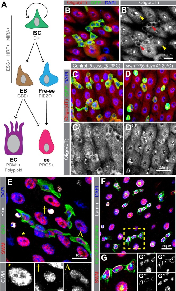
Fig. 1.

Knockdown of swm in intestinal progenitor cells leads poly(A)+ RNA accumulation in nuclei. a) A schematic of intestinal cell types and markers. In this study, we refer to ESG+ cells as progenitor cells (Ps), which include both ISCs and EBs. b) Poly(A)+ RNA distribution in cells from esgTS intestines, labeled by oligo (dT) probes (red) and Ps are labeled with GFP (anti-GFP in green) and nuclei with DAPI (blue). b′) Poly(A)+ RNA distribution is shown in gray scale in Ps (red arrowheads) and ECs (yellow arrowheads). Sections from (c) esgTS and (d) esgTS; swmRNAi PMGs after 5 days at 29°C stained with oligo (dT) (red), GFP (green), and DAPI (blue). e) PMG section from esgTS stained for Swm (anti-Swm in red). Ps are shown in GFP (green), ees are stained for Prospero (anti-Prospero in white) and nuclei for DAPI (blue). Enlarged EC (*), ee (†) and P (Δ) show Swm in gray scale. Individual channels of (e) are shown in Supplementary Fig. 1. (f and g) PMG region from UAS-myr GFP driven by esg-GAL4 stained for Swm (anti-Swm in red) and counterstained for cell membrane of Ps (myr-GFP in green), nucleoli of all cells (anti-Fibrillarin, nuclear green), nuclear membranes of all cells (anti-Lamin in white), and nuclei (DAPI in blue). (g) Enlargement of cells indicated in (e) with individual channels for Swm in red (g′), cell membranes (myr-GFP) and nucleoli (Fibrillarin) in green (g″), nuclear membranes in white (g′′′) and nuclei in blue (g′′′′). Complete genotypes are listed in Supplementary Table 1. P, progenitor cell; EC, enterocyte; ee, enteroendocrine cell; Pros, Prospero; PMG, posterior midgut.
