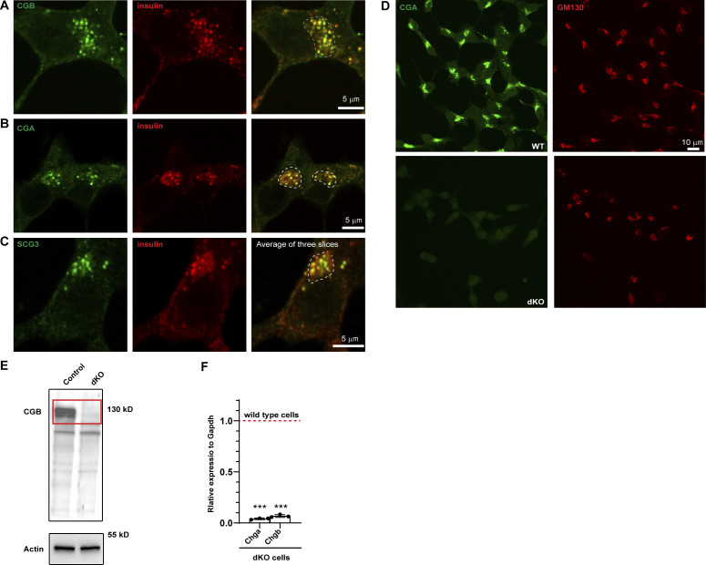
Figure S3.
Localization of endogenous granin proteins with insulin in INS1 832/13 cells and validation of CGA/CGB double KO cells. (A–C) INS1 832/13 cells fixed and labeled with antibodies to insulin in or CGB (A), CGB (B) and SCG III (C) in green. Insulin puncta at the Golgi apparatus in the perinuclear region, outlined using the dashed lines in the merge colocalize with each of the granin proteins. For CGB and CGA, images are a single slice from a confocal stack and in case of SCG III, an average projection from three consecutive slices from a confocal image. (D) Representative images of INS1 832/13 cells (wild type; top) and CGA/CGB double knockout (dKO; bottom) stained for CGA and GM130 antibodies to validate absence of CGA staining from the dKO cells. (E) Western blot (top) shows cell lysates from INS1 832/13 wild-type and dKO cells probed using CGB antibody. Note the absence of band in dKO cells which have been highlighted using the red rectangle. The bottom blot is probing of the same membrane for actin, which is used as a loading control. (F) qPCR to monitor the reduction in transcripts for Chga and Chgb in wild-type and dKO cells. Values are represented as mean ± SD from three independent experiments and expressed relative to Gapdh. Relative values for wild-type cells are normalized to 1. Statistical analysis was performed using unpaired two-tailed t test ***P < 0.001. Source data are available for this figure: SourceData FS3.

