Figure 5.

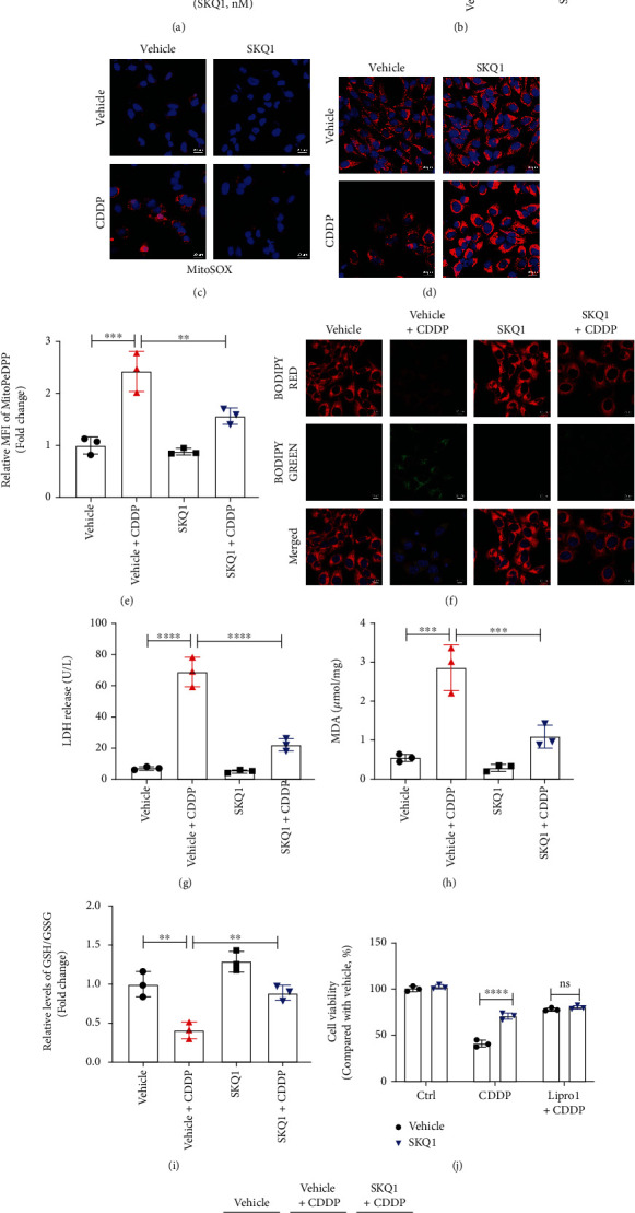
SKQ1 suppresses CDDP-induced mitochondrial oxidative stress and ferroptosis in HK2 cells. (a) Viability of HK2 cells after treatment with various concentrations of SKQ1. (b) The relative MitoSOX levels of HK2 cells treated with CDDP (10 μg/mL) with or without 20 nM SKQ1 were then analyzed by flow cytometry. Representative fluorescence images of (c) MitoSOX and (d) TMRM in the four different groups of HK2 cells (red, MitoSOX or TMRM; blue, Hoechst). (e) Mitochondrial lipid peroxidation was evaluated by staining with MitoPeDPP and MFI of oxidized MitoPeDPP and analyzed using flow cytometry. (f) Representative fluorescence images of lipid peroxidation after cell staining with BODIPY dye (red, lipids; green, oxidized lipids; blue, Hoechst). (g) LDH release from HK2 cells for all groups, and (h) relative levels of MDA and the (i) relative GSH/GSSG ratio in HK2 cells treated with CDDP and cotreated with vehicle or SKQ1. (j) We applied a CCK-8 assay to ascertain the viability of HK2 cells treated with SKQ1, 10 μM liproxstatin-1, or 20 mM Z-VAD-FMK, respectively, or with a combination of all three, followed by treatment with CDDP. (k) Immunoblots of GPX4 and cleaved caspase-3 in HK2 cells after treatment with CDDP and cotreatment with vehicle or SKQ1. Three independent assays were conducted. ∗∗∗∗P < 0.0001, ∗∗∗P < 0.001, ∗∗P < 0.01, and ∗P < 0.05 (by one-way or two-way ANOVA).
