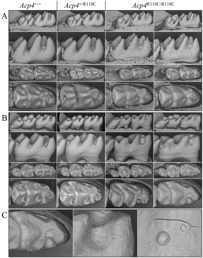
Figure 6.
Backscattered SEM Images of Acp4+/+, Acp4+/R110C, and Acp4R110C/R110C Mandibular Molars from D14 and 7-week Mice. (A) At D14 the mandibular first molars are very close to erupting into the oral cavity. The soft tissues have been removed to see the completed crown surface prior to potential post-eruptive changes. (B) The 7-week molars have been in function for about 5 weeks and feeding on a soft chow diet. (C) Close views of a 7-week Acp4R110C/R110C mandibular first molar. The Acp4+/+ and Acp4+/R110C molars are indistinguishable. The cusps of D14 Acp4R110C/R110C molars appear slender and pointed as the mineralized material covering them is much thinner than normal enamel. This layer of aplastic enamel is studded with small nodules that give the surface a rough appearance. The 7-week Acp4R110C/R110C molars show severe attrition and dentin exposure due to the absence of a true enamel layer. Most mineralized nodules that appeared at D14 have been abraded or chipped away, while some remain on the tooth surface.

