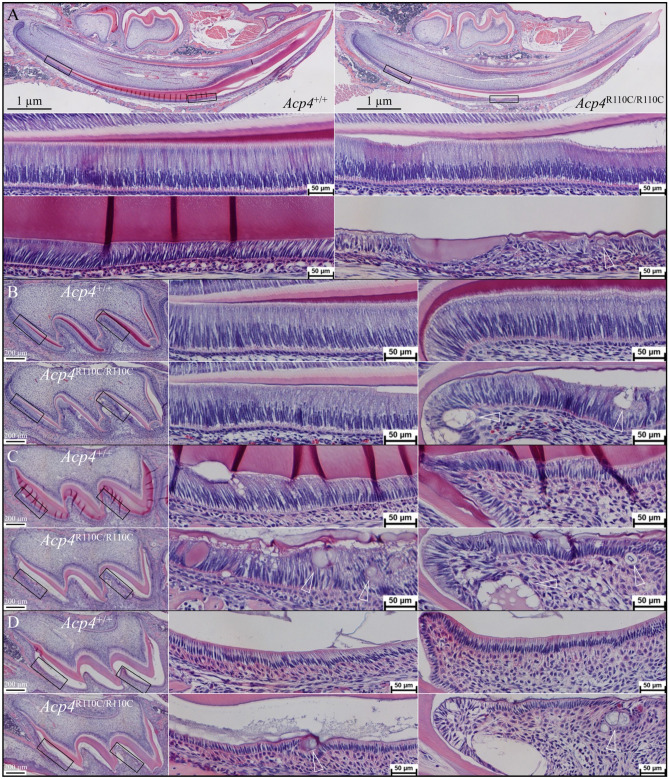
Figure 8.
Histology of Sagittally Sectioned, H&E Stained, Developing Incisors and Molars from Acp4+/+ and Acp4R110C/R110C Mice. (A) Histology of D12 mandibular incisors. Top: sections through Acp4+/+ (left) and Acp4R110C/R110C (right) hemimandibles (40×). Black rectangles outline positions of higher magnification (200×) images shown below, covering the initiation of enamel formation (middle) and post-secretory transition (bottom). Note that in Acp4R110C/R110C mice no proper enamel matrix is formed. Ameloblasts detach but remain polarized during the secretory stage (middle), but progressively degenerate as pathological ectopic mineral nodules (arrowhead) form in the soft tissue. (B–D) Histology of D4 (B), D8 (C), and D12 (D) Acp4+/+ and Acp4R110C/R110C maxillary first molars. Left: whole molar sections with black rectangles outlining ameloblasts on the mesial and distal cusps shown at higher magnification (200×) toward the right. As in the incisors, in Acp4R110C/R110C molars no proper enamel formation was observed. Ameloblasts polarized but detached from the mineral surface, ectopic mineral nodules formed (arrowheads) and expanded, and cell pathology worsened with time.

