Fig. 3.
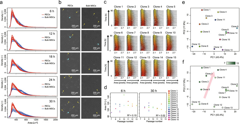
Morphological characterization of 15 RECs. a Size distribution comparison between RECs (15 clones) and conventionally processed bulk MSCs (cMSCs, 9 lots) and their time-course changes. Only adherent and extending cells are counted. The dotted vertical line represents the average cell sizes. b Representative time-course images of REC and Bulk MSC. Yellow and blue arrows represent proliferating cells and proliferated cells in near time, respectively. c Size distribution and their time-course changes among 15 clones. d Correlation of “SD of area” and serial-passage limitation number. R2 indicates the coefficient of determination. e, f PCA plot of 15 RECs profiled by 24 morphological descriptors. PCA plot with clone color labels (e). PCA plot colored by the heatmap of their serial-passage potencies (f)

