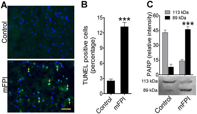
Fig. 7.
Activation of caspase-1 induces intrinsic apoptosis in fluid percussion-injured rats: a TUNEL staining (green) of rat cortex tissue sections without injury (control) and 24 h after FPI. Arrows indicate TUNEL-positive cells. The nucleus was counterstained with DAPI (blue). Scale bar 100 μm. b Percentage of TUNEL-positive cells in the adjacent area of FPI and compared with control. Values are mean ± SEM (n = 6); ***p < 0.001. c Western blotting of PARP protein levels in rat brain cortex lysates in control and 24 h after FPI. Bar graphs show the changes in relative intensity of 113- and 89-kDa fragments of PARP. Values are mean ± SEM (n = 6); ***p < 0.001

