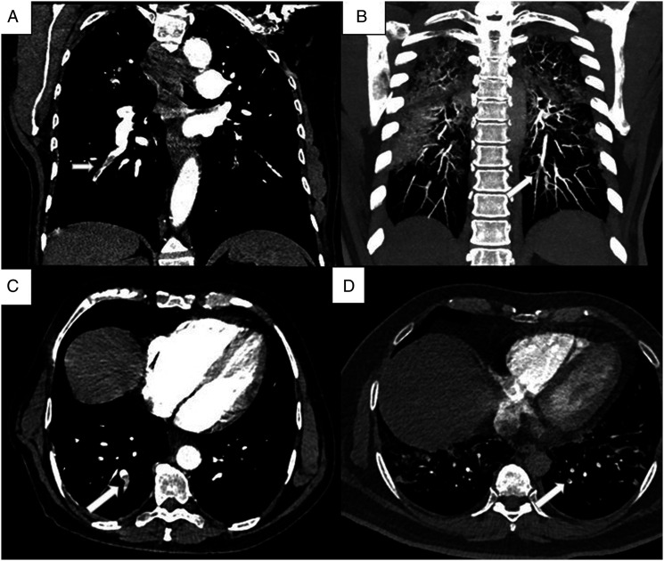
Figure 3.
(A - D): CTPA images of a 54-year-old male with respiratory distress. Coronal (A) and axial (C) CT images show hypodense filling defect consistent with thrombus (arrow) in the segmental and subsegmental branches of right lower lobe. CTPA images of a 38-year-old male with elevated d-dimer value. Coronal maximum intensity projection (B) and axial (D) images show thrombi in the subsegmental branches (arrow).

